 Skip to content

An Achilles tendon injury in dogs, more precisely an injury to the common calcaneal tendon complex, can allow the hock to collapse into an abnormal flexed or flat-footed position. The change may appear suddenly after trauma or develop against a background of chronic tendon degeneration. Either pattern needs veterinary assessment because a similar-looking “dropped hock” can arise from a different structure and require a different treatment.
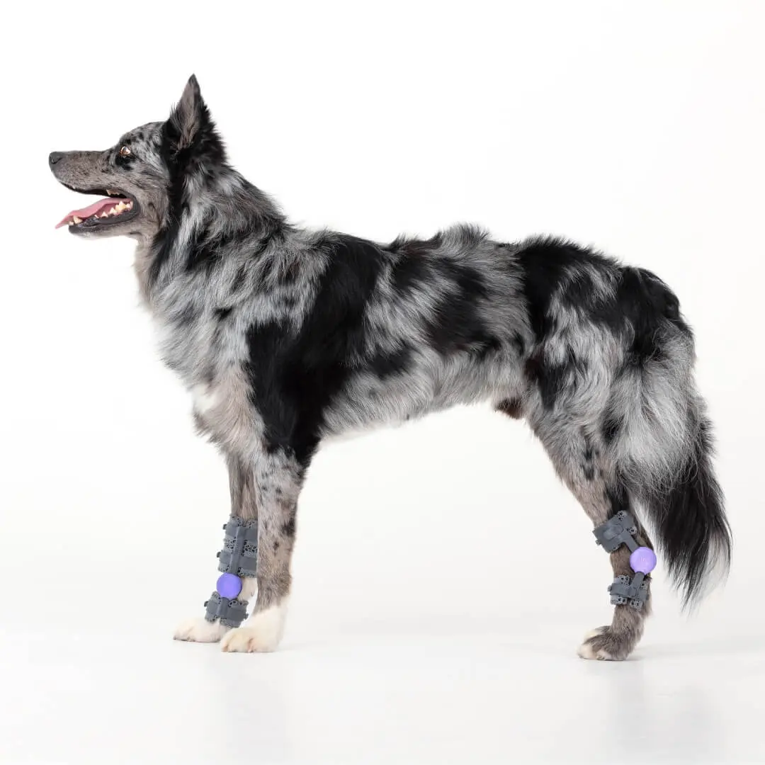
This guide explains the anatomy, warning signs, diagnostic process, surgical and conservative options, and the carefully defined role of external support. A hinged tarsal orthosis can help manage prescribed motion in selected cases, but it cannot reconnect a ruptured tendon or replace the protection required during early healing.
What Is an Achilles Tendon Injury in Dogs?
The structure commonly called the canine Achilles tendon is not one cord. It is a complex formed by the gastrocnemius tendon, superficial digital flexor tendon, and the combined tendons of the gracilis, semitendinosus, and biceps femoris muscles. Together, these components insert at or around the calcaneus and help maintain normal tarsal extension during weight-bearing.[1]
A common calcaneal tendon injury may affect one component, several components, or the complete complex. When the system can no longer resist flexion normally, the hock sinks toward the ground during stance. This mechanical effect is central to understanding an Achilles tendon injury in dogs and to selecting any form of immobilisation, surgery, rehabilitation, or external support.
Key terminology: “Achilles tendon” is the familiar owner-facing term. “Common calcaneal tendon complex” is more anatomically precise because several tendons contribute to the mechanism. The hock and tarsus refer to the same joint region in this article.
Why a Dropped Hock Is Not a Diagnosis
Owners may describe both tendon failure and ligament instability as a hock that “drops,” but the abnormal movement is different. In an Achilles tendon injury in dogs, loss of tendon function usually permits excessive tarsal flexion and may create a plantigrade stance. In structural tarsal hyperextension, failure of plantar supporting structures permits excessive extension. A device configured to limit the wrong direction of movement would not match the lesion.
Fracture, luxation, collateral ligament injury, neurological weakness, and compensation for disease elsewhere in the limb can also alter hock position. This is why a photograph, symptom list, or online sizing chart cannot determine whether a custom tarsal orthosis is suitable. For the broader differential diagnosis, see WIMBA’s guide to tarsal instability in dogs.
Partial Injury, Complete Rupture, and the Crab-Claw Posture
The clinical picture depends on which components remain functional. A complete disruption can produce marked hock flexion and a flat-footed or plantigrade posture. In a partial canine Achilles tendon injury, the superficial digital flexor tendon may remain intact while the gastrocnemius and combined tendon are damaged. Its continued pull can curl the toes into the characteristic “crab-claw” position.[2]
The posture is a useful clue, not a complete diagnosis. The tendon may be injured at its attachment to bone, within the tendon body, or near the musculotendinous junction. A veterinarian must determine the site, chronicity, severity, and tissues involved before recommending treatment for an Achilles tendon injury in dogs.
What Causes Achilles Tendon Injury in Dogs?
An acute Achilles tendon injury in dogs may follow a laceration, collision, fall, severe stretch, or other trauma. Chronic or atraumatic cases may involve progressive degeneration, repeated micro-injury, or an underlying systemic factor. The American College of Veterinary Surgeons notes associations with conditions such as hypothyroidism, hyperadrenocorticism, and immune-mediated polyarthritis, but an individual dog still requires its own diagnostic workup.[1]
In a retrospective ultrasound series of 43 dogs, 39 injuries were partial and four involved complete disruption of all three components. Abnormalities were bilateral at presentation in 21 dogs, and four of the 22 initially unilateral cases later developed changes in the opposite tendon.[2] These findings do not predict what will happen to every patient, but they support examination and monitoring of both hind limbs after a common calcaneal tendon injury.
Signs That Need Veterinary Assessment
The signs of an Achilles tendon injury in dogs vary with severity and duration. Observe the dog standing, rising, walking, and turning on a non-slip surface. Short videos from the side and behind can help the veterinary team see how the posture changes under load.
- A flat-footed or plantigrade stance with the metatarsus closer to the floor
- Toes curled downward into a crab-claw posture
- Swelling, heat, thickening, bruising, or sensitivity behind the hock
- Limping, toe-touching, or inability to bear normal weight
- A shortened stride, weak push-off, or reduced propulsion
- Difficulty rising, using stairs, jumping, or maintaining usual activity
- A sudden change after trauma or a progressive change over several weeks
When to Seek Prompt Care
Seek prompt veterinary assessment if the dog cannot bear weight, the hock suddenly collapses, an open wound lies near the tendon, swelling increases rapidly, pain is severe, or the paw becomes cold or discoloured. Urgent care for an Achilles tendon injury in dogs starts with protecting the limb from further trauma. Do not repeatedly flex, extend, press, or twist the hock to test it. Do not improvise a rigid splint or purchase a brace before the damaged structure is identified.
How Achilles Tendon Injury in Dogs Is Diagnosed
Assessment of an Achilles tendon injury in dogs begins with the history and a complete orthopaedic examination. The clinician evaluates stance, gait, pain, swelling, tendon continuity, toe position, active and passive movement, muscle condition, skin, and the opposite limb. A suspected common calcaneal tendon injury may also prompt a neurological examination when weakness or altered paw placement could contribute to the posture.
Musculoskeletal ultrasound can show fibre-pattern disruption, thickening, irregularity, and the components and region affected by a canine Achilles tendon injury. In the 43-dog series, ultrasound was used to classify lesions and correlate findings with surgery where available.[2] The test is operator-dependent, so equipment, technique, and experience matter.
Radiographs help assess the calcaneus, mineralisation, osteoarthritis, alignment, and possible avulsion or concurrent fracture. CT or MRI may be selected for complex bone, joint, tendon, ligament, or soft-tissue questions when the result could change management. The goal is not merely to label an Achilles tendon injury in dogs. It is to define what failed and what must be protected.
Treatment Depends on the Lesion and Healing Stage
There is no single protocol for every Achilles tendon injury in dogs. The plan may combine activity restriction, veterinary pain management, surgical repair, temporary immobilisation, external orthotic support, staged rehabilitation, and scheduled imaging or gait reassessment.
- Surgical tendon repair: commonly considered for complete disruption, major avulsion, severe partial injury, or a functionally important gap
- Temporary protection: a cast, splint, transarticular fixation, or another surgeon-selected method may protect a repair during vulnerable early healing
- Pantarsal arthrodesis: may be selected as a primary or salvage procedure in some chronic, severe, or failed cases
- Conservative management: may be considered for selected stable partial injuries when close monitoring and owner compliance are realistic
- Rehabilitation: progresses only as the treating team confirms that healing tissue can tolerate the planned load and range
A 2026 multicentre retrospective study evaluated 89 surgeries in 80 dogs with common calcaneal tendon pathology. Overall complications occurred in 46 of 89 limbs. Tendon repair with temporary tarsocrural immobilisation had a higher catastrophic complication rate than pantarsal arthrodesis in that cohort.[3] At the final in-clinic follow-up (median 10 weeks), 64 of 89 limbs (72%) had full or acceptable function, although follow-up was shorter than 12 weeks for most dogs.[3] The groups were not randomised, and the finding does not make one operation correct for every common calcaneal tendon injury. It does support a detailed discussion of revision risk, aftercare, cost, and expected function.
Why Early Immobilisation and Later Orthotic Support Are Not Interchangeable
A freshly repaired tendon may require strict protection against gapping. A hinged custom tarsal orthosis intended for controlled walking later in recovery should not be assumed to provide the same protection as a cast, splint, temporary screw, transarticular plate, or external skeletal fixator. The surgeon determines when and how the patient moves from one stage to the next.
Biomechanical work in six dogs also found that immobilising the tarsal joint did not eliminate common calcaneal tendon strain during weight-bearing.[4] This helps explain why an external hock device should not be described as completely unloading an Achilles tendon injury in dogs. External support can change motion and loading, but biological healing and controlled activity remain central.
What the Evidence Shows About Orthotic Support
Published clinical evidence for orthotic management of an Achilles tendon injury in dogs is limited. One case report described a dog with gastrocnemius tendon strain treated with autologous mesenchymal stem cells and a progressive custom orthosis. Clinical and force-platform measures improved, but a single patient receiving combined interventions cannot establish how much benefit came from the orthosis alone.[5]
A broader prospective study followed 43 dogs using custom orthoses or limb-replacement devices. Thirty-nine experienced at least one complication, with skin problems common during the first three months.[6] This was not an Achilles-specific trial, but it supports gradual introduction, professional fitting, and planned rechecks whenever a custom tarsal orthosis is used.
Current evidence therefore supports cautious, case-specific use rather than a promise of tendon healing or avoidance of surgery. An orthosis may help control a prescribed movement while worn. It does not regenerate tendon fibres, guarantee a normal gait, or remove the need for diagnostics and follow-up.
When WIMBA Tarsus Orthosis GO May Be Considered
Current WIMBA information lists Tarsus Orthosis GO for eligible mild sagittal-plane instability and tarsal hyperflexion associated with Achilles or common calcaneal tendon injuries when no complete tear is present. Hyperflexion cases require clinician-selected custom Tokens to limit flexion.[7] This means an Achilles tendon injury in dogs does not automatically qualify for GO.
For an Achilles tendon injury in dogs, a veterinarian or rehabilitation professional must decide whether the lesion is incomplete, whether sagittal-plane control matches the problem, and whether the current stage permits therapeutic activity. The intended role may be controlled support during prescribed walks or rehabilitation. GO is not intended for acute postoperative use, strict immobilisation, sprinting, agility, or repetitive jumping.
A focused WIMBA case review should establish:
- Whether the tendon injury is partial or complete
- Which components and anatomical level are affected
- Whether instability is limited to the sagittal plane
- Which flexion limit and activity the device should support
- Whether wounds, swelling, skin disease, anatomy, body weight, or behaviour affect candidacy
- How the outcome and stopping criteria will be measured
More advanced, multiplanar, or otherwise complex instability requires separate review and may call for a PRO configuration, surgery, or another pathway. WIMBA does not offer a one-device answer for every Achilles tendon injury in dogs. Clinics should use the latest WIMBA Tarsus Orthosis information and submit uncertain cases before ordering.
How the GO Pathway Is Measured and Ordered
For an eligible GO case involving an Achilles tendon injury in dogs, the clinic collects two required limb photographs and product-specific manual measurements through WimbaAPP. WimbaSCAN is not required for this pathway. The clinical team also needs the diagnosis, relevant history, skin and wound status, intended functional goal, and the prescribed motion-control configuration.
- Diagnose the lesion: identify the components, severity, chronicity, plane of instability, and healing stage.
- Confirm candidacy: compare the patient with current GO indications and submit a case when the situation is uncertain or complex.
- Collect patient data: take the required photographs and guided measurements according to the current WimbaAPP workflow.
- Define motion control: select the prescribed flexion limit rather than applying a default Token setting.
- Fit dynamically: assess alignment, retention, skin clearance, joint control, and gait before home use.
- Reassess: review fit, healing, motion settings, skin, gait, and the original clinical goal.
The result is a patient-specific custom tarsal orthosis, not an off-the-shelf sleeve selected by circumference alone. Custom manufacture can improve anatomical matching, but it does not eliminate migration, pressure, or tolerance problems. The first fitting remains a clinical appointment.
First Fitting and Gradual Introduction
At fitting, the hinges should sit at the tarsal joint, the frame should remain correctly aligned, and straps should apply even tension. No hard component should contact the limb through the gait cycle. The device should not rotate, twist, or slide during controlled walking. For a healing Achilles tendon injury in dogs, the clinician must also check that the prescribed flexion limit is functioning as intended.
Current WIMBA Tarsus Orthosis GO guidance starts with up to 15 minutes of controlled leash walking, no more than three times daily during the initial period. Wearing time may then increase by 15 minutes every two days when skin, gait, behaviour, and device position remain acceptable. The treating veterinarian or therapist may prescribe a different schedule for a canine Achilles tendon injury.[8]
The orthosis is not intended for continuous 24-hour wear, and the dog must remain supervised. A provider may recommend a thin breathable layer or correctly applied veterinary wrap, but an incorrectly placed wrap can itself cause swelling or injury. Owners should use the method demonstrated by the clinic.
Skin, Gait, and Device Monitoring
Inspect the skin after every session during introduction, paying particular attention to the tendon region, bony prominences, dewclaw, strap edges, and any area affected by changing swelling. Remove the dog hock brace and contact the clinic if redness persists, hair is lost, skin breaks, swelling develops, the limb becomes painful, or gait worsens.
Check the frame, hinges, Tokens, fasteners, straps, and inserts before use. Do not heat, cut, drill, grind, bend, or reshape the structure. Do not change the range of motion without professional direction. Hand-clean the device with lukewarm water and mild soap, then allow it to air-dry according to the manual.[8]
Monitoring an Achilles tendon injury in dogs must account for a changing limb. Swelling may decrease, muscle mass may change, the tendon may thicken, and activity may progress. A well-fitted device can therefore require adjustment or reassessment over time. Do not compensate for movement by simply tightening every strap.
Rehabilitation Must Follow Tissue Healing
Rehabilitation after an Achilles tendon injury in dogs is staged, not calendar-driven alone. Early priorities may include protecting the repair, controlling pain and swelling, maintaining safe mobility, and preventing avoidable complications. Later phases may introduce clinician-selected range-of-motion work, controlled weight-bearing, muscle recruitment, balance, and gradual endurance.
The surgeon or rehabilitation professional decides when each activity is safe. Underwater treadmill work, cavaletti, inclines, strengthening, and longer walks are not automatically appropriate simply because a device is present. A dog hock brace can support a defined exercise, but it does not make unrestricted running, jumping, or rough play safe.
Useful outcomes for an Achilles tendon injury in dogs include improved hock position during stance, better controlled push-off, reduced lameness, increased tolerance of prescribed activity, stable skin, and imaging or clinical evidence that supports progression. If the dog becomes more lame, shortens the stride, rotates the limb, or resists an activity, pause and request reassessment rather than pushing through the change.
Questions to Ask the Veterinary Team
- Is this a partial or complete injury, and which tendon components are affected?
- Is the abnormal posture caused by tendon injury, ligament failure, bone injury, or another condition?
- Does the lesion require surgery or strict immobilisation?
- At what healing stage could controlled motion or external support begin?
- Would a custom tarsal orthosis match the plane and severity of instability?
- Which range, activity, wear schedule, and rehabilitation exercises are prescribed?
- Which findings require an urgent call or an earlier recheck?
These questions turn treatment of an Achilles tendon injury in dogs into a defined plan with measurable checkpoints. They also help separate the role of biological healing, surgery, temporary protection, rehabilitation, and orthotic support.
Start With a Veterinary Hock Assessment
A dropped hock can reflect several different injuries. Veterinary professionals can submit the diagnosis, imaging, history, and intended functional goal for case review. Pet owners can find a WIMBA Provider for examination and clinician-led fitting.
Frequently Asked Questions About Achilles Tendon Injury in Dogs
These answers provide general guidance. They cannot diagnose a tendon lesion, determine surgical candidacy, or confirm whether an individual patient can safely use an orthosis.
What is an Achilles tendon injury in dogs?
An Achilles tendon injury in dogs is damage to one or more components of the common calcaneal tendon complex behind the hock. The injury can reduce the limb’s ability to maintain normal tarsal extension, creating lameness, swelling, excessive hock flexion, a flat-footed stance, or curled toes.
Is a canine Achilles tendon injury the same as tarsal hyperextension?
No. A canine Achilles tendon injury usually permits excessive tarsal flexion because the tendon mechanism cannot maintain extension. Structural tarsal hyperextension usually reflects failure of plantar supporting structures and permits excessive extension. Examination and imaging are needed because the required treatment and motion control differ.
Can a dog recover from a common calcaneal tendon injury without surgery?
Selected stable partial injuries may be managed conservatively under close veterinary direction. Complete disruption, major avulsion, or severe functional instability commonly prompts surgical assessment. The decision depends on the damaged components, gap, chronicity, tissue quality, posture, patient factors, and realistic aftercare.
Can a dog hock brace heal a torn Achilles tendon?
No. A dog hock brace can limit a prescribed range while worn, but it cannot reconnect a complete rupture or replace surgical repair when repair is indicated. It may be one component of conservative management or later rehabilitation for an eligible partial injury.
How is an Achilles tendon injury in dogs diagnosed?
Diagnosis combines history, orthopaedic examination, stance and gait assessment, and imaging. Ultrasound can assess tendon fibre pattern and identify affected components. Radiographs help evaluate the calcaneus, mineralisation, arthritis, and bone injury. CT or MRI may be used for selected complex cases.
When may WIMBA Tarsus Orthosis GO be considered?
Current WIMBA information includes eligible mild sagittal-plane cases and tarsal hyperflexion associated with an incomplete tendon injury. The clinician must confirm there is no complete tear, define the flexion limit, and determine that therapeutic activity is appropriate. Acute postoperative protection and strict immobilisation require a different pathway.
How long should a dog wear a tarsal orthosis?
The schedule is individual. Current GO guidance begins with controlled sessions of up to 15 minutes, no more than three times daily, followed by gradual increases when skin, gait, behaviour, and device position remain acceptable. The orthosis is not intended for 24-hour wear, and the patient must remain supervised.
Medical Disclaimer: All information on this website is intended for instruction and information purposes only. The authors are not responsible for any harm or injury that may result. Significant injury risk is possible if you do not seek suitable professional advice about your patient’s specific situation. No guarantees of specific results are expressly made or implied on this website.
Scientific and Clinical References
- American College of Veterinary Surgeons. Achilles’ Tendon Injuries. Clinical resource. View clinical resource
- Gamble LJ, Canapp DA, Canapp SO. Evaluation of Achilles tendon injuries with findings from diagnostic musculoskeletal ultrasound in canines: 43 cases. Vet Evid. 2017, 2(3). doi:10.18849/ve.v2i3.92
- Wylie S, Piana F, Montgomery P, et al. Complications and outcomes following surgical management of common calcaneal tendon pathology in 80 dogs. Vet Surg. 2026, 55(2):437-447. doi:10.1111/vsu.70053
- Lister SA, Renberg WC, Roush JK. Efficacy of immobilization of the tarsal joint to alleviate strain on the common calcaneal tendon in dogs. Am J Vet Res. 2009, 70(1):134-140. doi:10.2460/ajvr.70.1.134
- Case JB, Palmer R, Valdes-Martinez A, Egger EL, Haussler KK. Gastrocnemius tendon strain in a dog treated with autologous mesenchymal stem cells and a custom orthosis. Vet Surg. 2013, 42(4):355-360. doi:10.1111/j.1532-950X.2013.12007.x
- Rosen S, Duerr FM, Elam LH. Prospective evaluation of complications associated with orthosis and prosthesis use in canine patients. Front Vet Sci. 2022, 9:892662. doi:10.3389/fvets.2022.892662
- WIMBA. Tarsal Orthotics for Dogs: Clinical Indications and Ordering Pathway. View current product information
- WIMBA Knowledge Base. WIMBA Tarsus Orthosis GO User Manual. Updated May 26, 2026. View current user manual
Related reading: Custom Dog Hock Orthotics for Daily Walking Support · 3D-Printed Dog Hind Limb Orthotics: Fit, Use, and Evidence