 Skip to content

Tarsal Instability in Dogs: Causes, Signs, and Support Options
Tarsal instability in dogs can make the hock look lower, straighter, more flexed, or angled to one side when a dog stands and walks. The appearance may be dramatic, but it does not identify which ligament, tendon, joint row, or nearby structure is responsible.
This guide explains the main patterns of tarsal instability in dogs, why tarsal hyperextension and common calcaneal tendon injury are not the same problem, how veterinarians investigate dog hock instability, and when external support such as a custom dog tarsal brace may fit into a wider treatment plan.
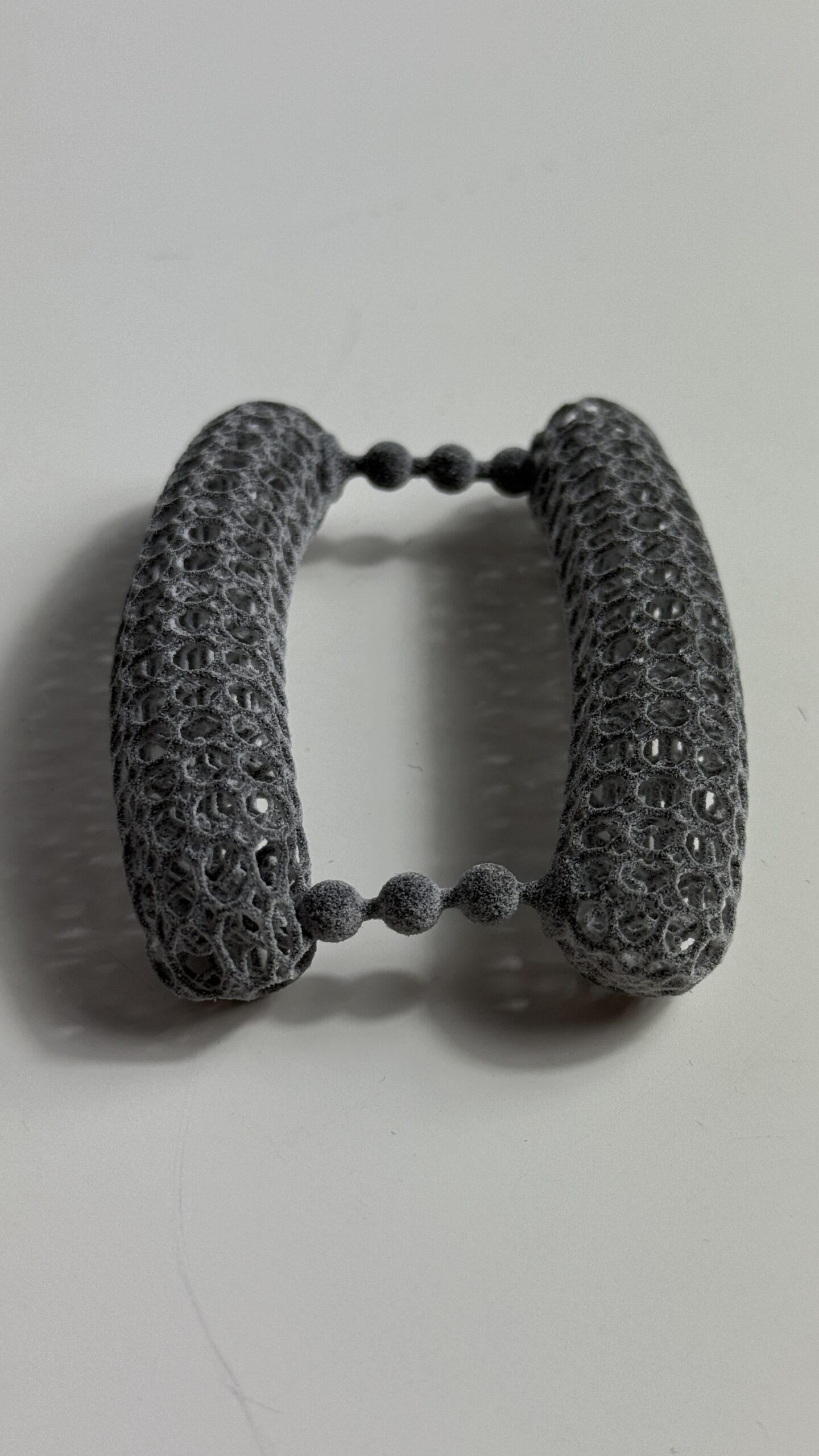
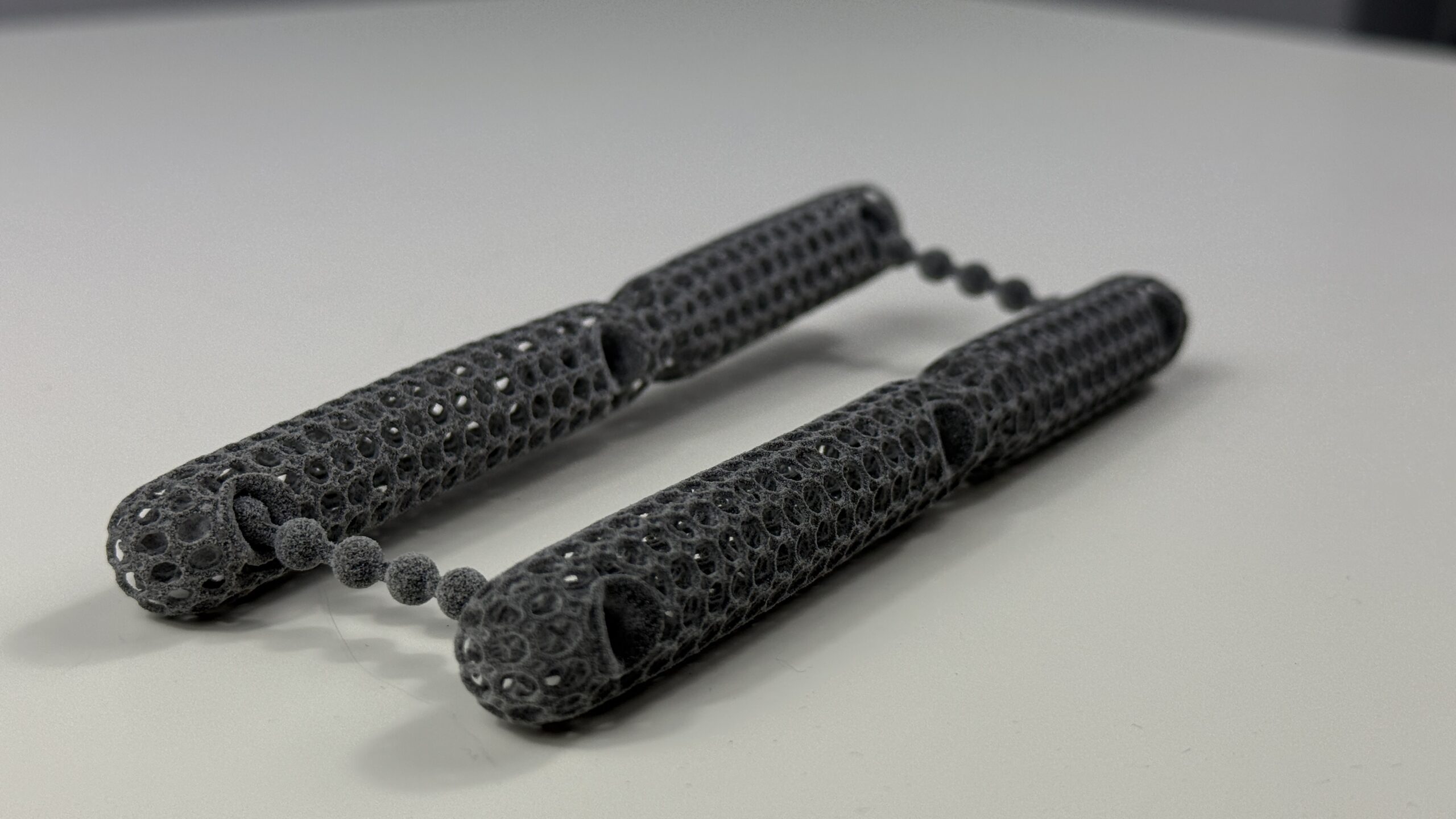
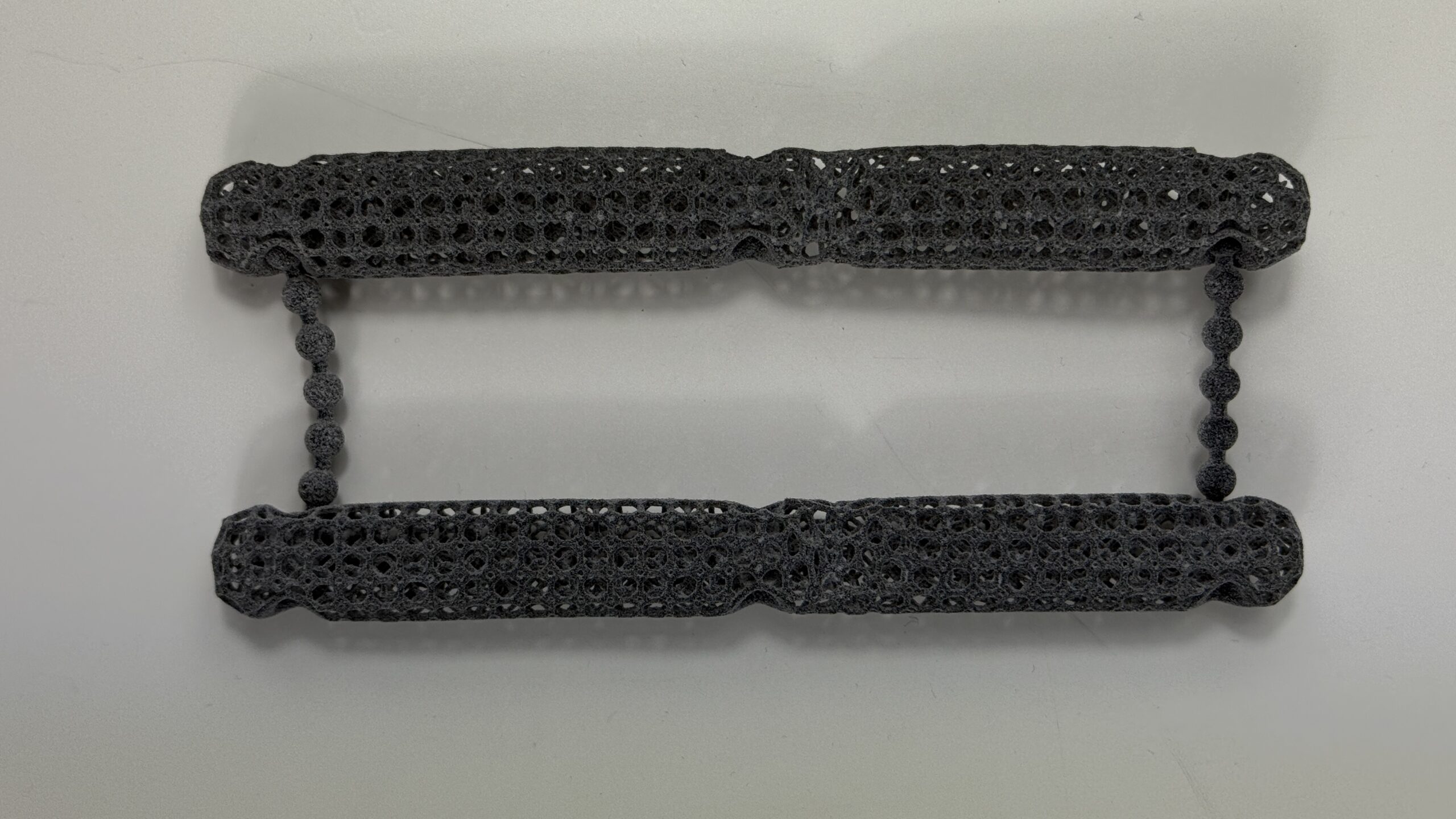
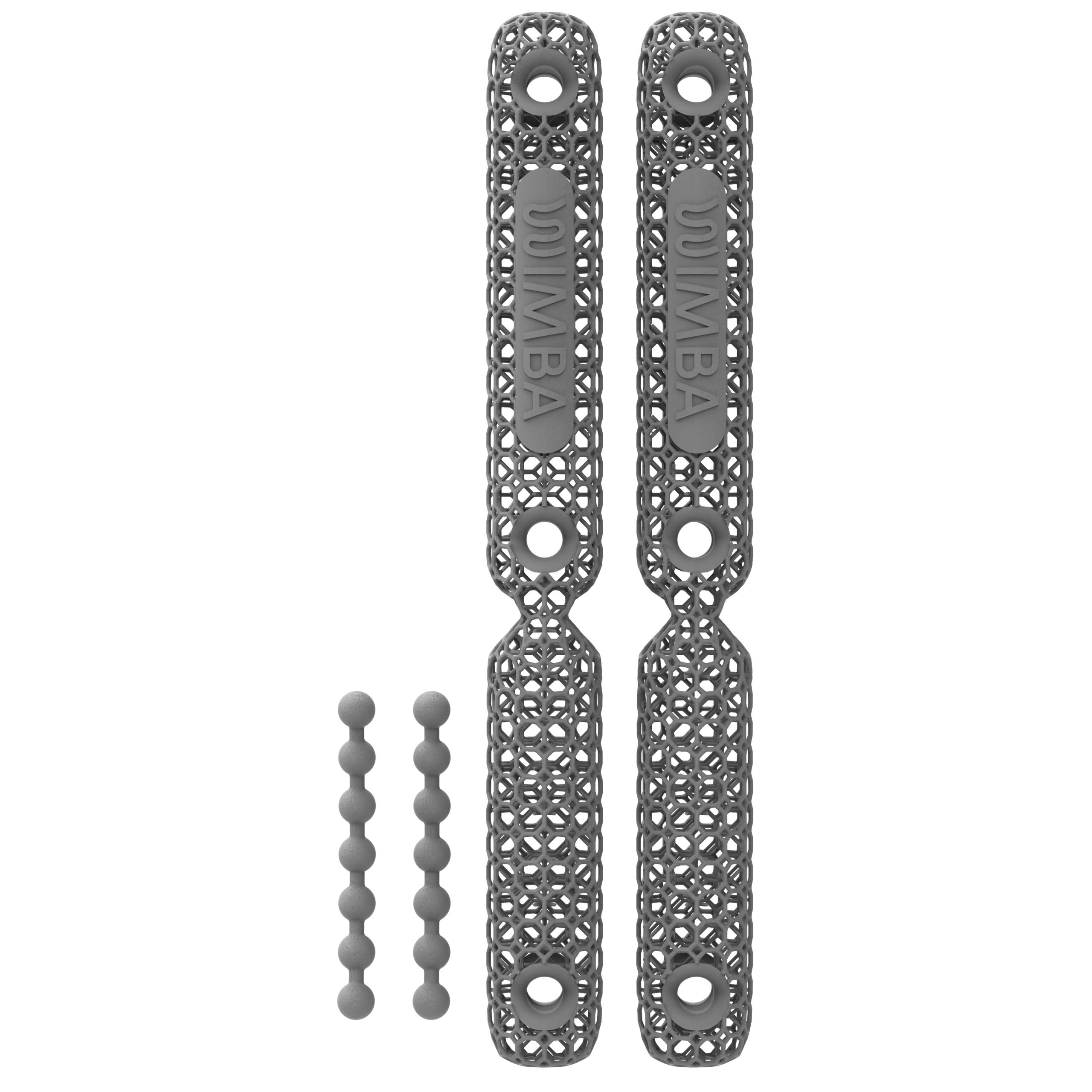
Non-surgical management of tarsal instability in dogs may combine activity restriction, veterinary pain management, rehabilitation, weight management, and external orthotic support, depending on which structure is affected. WIMBA makes the WIMBA Tarsus Orthosis GO, a custom 3D-printed tarsal orthosis for dogs and cats that supports the hock during walking in selected cases of tarsal hyperextension or instability. It is prescribed and fitted through veterinary professionals, and it does not replace surgery when a tendon, ligament, or fracture needs repair.
A “dropped hock” is a sign, not a diagnosis
Failure of the plantar supporting ligaments, injury to the common calcaneal tendon complex, collateral ligament damage, fracture or luxation, neurological dysfunction, and a compensatory gait pattern can produce different forms of abnormal tarsal posture. Do not try to identify the cause by forcing the joint through its range at home.
What Is Tarsal Instability in Dogs?
The tarsus is the complex joint region between the lower leg and the metatarsus in a dog’s hind limb. Owners commonly call it the ankle or hock. It contains seven tarsal bones arranged across several joint levels, supported by joint capsules, collateral and plantar ligaments, the common calcaneal tendon complex, and surrounding muscles and tendons.12
Tarsal instability in dogs means that one or more of those structures no longer controls position or motion normally. The instability may occur mainly in the sagittal plane, producing excessive flexion or extension, or in a medial-lateral direction, producing varus, valgus, rotation, or subluxation. It may be fixed, visible only during weight-bearing, or apparent only when a veterinarian performs a controlled examination.
| Observed pattern | What it may indicate | Important distinction |
|---|---|---|
| Excessive extension | Failure or laxity of plantar supporting structures, sometimes with intertarsal or tarsometatarsal injury | This is the pattern commonly described as tarsal hyperextension in dogs |
| Excessive flexion or plantigrade stance | Partial or complete common calcaneal tendon complex injury | The hock may look “dropped,” even though the mechanical problem is hyperflexion |
| Inward or outward deviation | Collateral ligament injury, luxation, fracture, or multiplanar instability | A simple sagittal-plane brace may not provide the required control |
| Weight-bearing-dependent posture | Structural instability, weakness, neurological dysfunction, or compensation from another joint | Visible posture alone cannot prove that the tarsus itself is the primary lesion |
Tarsal Hyperextension and Achilles Injury Are Different
The terminology can be confusing because owners may describe both conditions as a hock that “drops.” The direction of abnormal joint motion and the structure that has failed are clinically important.
Tarsal hyperextension
In structural tarsal hyperextension in dogs, plantar ligaments or other supporting structures fail to resist excessive extension during loading. Trauma can disrupt the plantar ligament, damage several ligaments, or occur together with fracture or luxation. Chronic calcaneoquartal instability has also been described, but the exact joint level and severity must be identified before treatment is chosen.1
Common calcaneal tendon injury
The structure commonly called the canine Achilles tendon is a complex formed by the gastrocnemius tendon, superficial digital flexor tendon, and the combined tendons of the gracilis, semitendinosus, and biceps femoris muscles. An Achilles tendon injury in dogs reduces the system’s ability to maintain normal tarsal extension, so the hock collapses into excessive flexion under load.3
A complete disruption may produce a flat-footed or plantigrade stance. In some partial injuries, the superficial digital flexor tendon remains functional and pulls the toes into a characteristic curled or “crab-claw” position. That posture is an important clue, but ultrasound or other imaging may still be needed to characterize which components are affected.34
What Causes Tarsal Instability in Dogs?
Tarsal instability in dogs is a descriptive finding rather than one disease. The following causes can require very different management.
Acute ligament injury, luxation, or fracture
A fall, collision, awkward landing, twisting injury, or shearing trauma can disrupt plantar or collateral ligaments and may also damage the tarsal bones or joint surfaces. Severe trauma can create instability in more than one direction. An apparently simple dog hock instability can therefore conceal a fracture, open injury, or luxation that requires urgent stabilization.
Common calcaneal tendon strain or rupture
An Achilles tendon injury in dogs may follow laceration, blunt trauma, severe stretching, or chronic degeneration. In a retrospective ultrasound series of 43 dogs, 39 had partial injuries and four had complete disruption of all three tendon components. Ultrasound abnormalities were bilateral at presentation in 21 of the 43 dogs, showing why examination of the opposite limb can matter.3
Collateral ligament injury
The medial and lateral collateral ligaments resist side-to-side deviation. Injury may create varus, valgus, or rotational instability and can occur with malleolar fracture or other tarsal trauma. Because the required control is not only flexion and extension, multiplanar tarsal instability in dogs needs different planning from mild sagittal laxity.
Osteoarthritis and chronic joint change
Osteoarthritis may develop after previous trauma or chronic abnormal motion and can add pain, stiffness, reduced range of motion, and periarticular thickening. It may also coexist with another cause of dog hock instability. Radiographic osteoarthritis does not by itself prove that joint laxity is the primary problem, so the treatment target still has to be defined.
Functional or compensatory posture
Not every hock that extends abnormally during stance has primary structural failure of the tarsus. A 2026 case report described bilateral, weight-bearing-dependent hyperextension associated with stifle pathology; the posture resolved after the stifles were treated without direct tarsal intervention.5 One case cannot establish a general rule, but it shows why the entire pelvic limb and neurological function should be assessed before labeling the problem as structural tarsal instability in dogs.
Signs of Tarsal Instability in Dogs
The signs of tarsal hyperextension in dogs and other hock disorders depend on the affected structure, chronicity, and whether the problem is unilateral or bilateral. Watch the dog standing, rising, walking, and turning on a non-slip surface rather than relying on one still image.
- A hock that sinks, opens excessively, or appears lower during weight-bearing
- A flat-footed or plantigrade stance with the metatarsus closer to the floor
- Toes curled downward in a “crab-claw” posture
- Inward or outward deviation of the hock or paw
- Limping, toe-touching, or reduced weight-bearing on the affected limb
- Swelling, heat, thickening, bruising, or sensitivity around the hock or tendon
- Shortened stride, weak push-off, difficulty rising, or reluctance to use stairs
- A change that is clearest during stance but less visible during the swing phase
- Repeated rubbing, licking, or skin injury near a prominent or collapsed joint
Short videos from the side and from behind can help the veterinary team see how tarsal instability in dogs changes with load. Do not repeatedly flex, extend, twist, or press the hock to test it. A painful or partially injured structure can be worsened by unsupervised manipulation.
When tarsal instability in dogs needs prompt care
Seek prompt veterinary assessment after significant trauma or when the dog cannot bear weight, the joint has suddenly collapsed, there is an open wound or obvious deformity, swelling is rapidly increasing, the paw becomes cold or discolored, pain is severe, or weakness and poor coordination are progressing. Do not apply a rigid splint or brace unless a veterinary professional has shown you how.
How Tarsal Instability in Dogs Is Diagnosed
Diagnosis begins with the history and a complete orthopedic examination. Because tarsal instability in dogs can originate at different joint levels, the veterinarian evaluates stance, gait, pain, swelling, active and passive motion, the level and direction of instability, muscle condition, skin, paw position, and the opposite limb. A neurological assessment may be added when weakness, altered paw placement, or a functional gait pattern is possible.
Because the canine tarsus contains multiple joint rows and closely associated soft tissues, examination alone may not localize every lesion. Imaging is chosen according to the suspected structure.2
Radiographs
X-rays evaluate bone alignment, fracture, luxation, mineralization, osteoarthritis, and other osseous change. Carefully performed stress views may be used when the clinician needs to document the direction and level of ligamentous instability.
Musculoskeletal ultrasound
Ultrasound allows direct assessment of tendon fiber pattern and can help identify the components and region affected in an Achilles tendon injury in dogs. Interpretation depends on equipment, technique, and operator experience.3
CT
Computed tomography provides detailed cross-sectional evaluation of complex fractures, bone fragments, articular involvement, and three-dimensional alignment when standard radiographs do not fully characterize the lesion.
MRI
Magnetic resonance imaging may be considered for selected tendon, ligament, muscle, or other soft-tissue questions. It is not required for every case and is chosen when the anticipated information could change management.
The diagnostic goal is not merely to confirm that the hock looks abnormal. It is to determine the structure, joint level, plane, severity, chronicity, and treatment objective behind tarsal instability in dogs.
Treatment for Tarsal Instability in Dogs Depends on the Structure
There is no single treatment protocol for tarsal instability in dogs. The plan may combine activity restriction, veterinary pain management, temporary immobilization, surgery, external orthotic support, rehabilitation, weight management, and scheduled imaging or gait reassessment.
Surgery or arthrodesis
Complete common calcaneal tendon disruption, severe plantar or collateral ligament failure, fracture-luxation, major multiplanar instability, or a chronically damaged joint may require tendon repair, ligament reconstruction, temporary internal or external stabilization, partial arthrodesis, or pantarsal arthrodesis. The chosen procedure depends on the lesion and specialist assessment.
A recent multicenter retrospective study reported outcomes and complications after 89 surgeries in 80 dogs with common calcaneal tendon pathology. It found substantial complication rates across procedures and a higher catastrophic complication rate after tendon repair with temporary tarsocrural immobilization than after pantarsal arthrodesis.6 This does not make one procedure correct for every patient; it shows why owners need a case-specific discussion of benefits, limitations, aftercare, and revision risk.
Selected non-surgical management
A veterinarian may consider conservative management for selected mild, stable, partial, or chronic presentations when the diagnosed tissue, patient health, functional goal, and expected owner adherence make that pathway reasonable. The plan may include controlled loading, external support, rehabilitation, and repeated examination or imaging. It should not be extrapolated to complete rupture, unstable fracture, or severe multiplanar collapse.
Postoperative protection and rehabilitation
After tendon or ligament surgery, the repair may require strict protection before controlled motion is introduced. Casts, splints, temporary screws, external skeletal fixation, or custom orthoses can serve different roles at different stages.4 The surgeon must decide when a dog tarsal brace is appropriate; an activity orthosis should not be assumed to provide the same immobilization as a postoperative construct.
Where a Dog Tarsal Brace May Fit
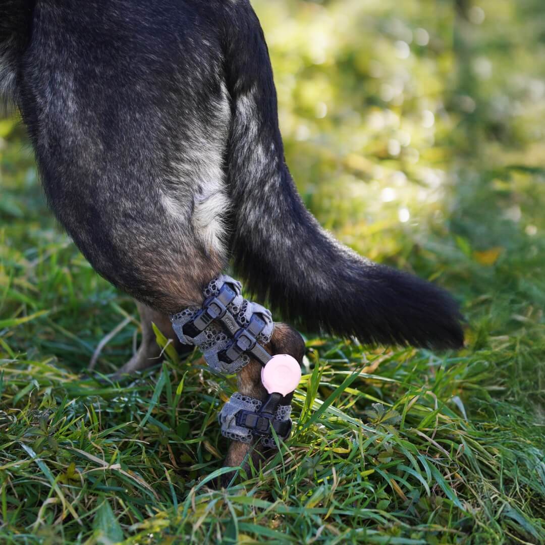
For selected tarsal instability in dogs, a custom dog tarsal brace is an external device designed to influence joint motion and provide support during activities defined by the treating professional. Depending on its geometry and range-of-motion settings, it may limit excessive extension, limit selected flexion, or help control more complex motion. It does not reconnect a ruptured tendon or ligament and cannot stabilize every fracture or luxation.
Biomechanical research also shows why a brace should not be described as completely unloading a healing tendon. In six dogs, immobilizing the tarsal joint did not eliminate common calcaneal tendon strain during weight-bearing.7 External control can change the mechanical environment, but muscle activity and loading still matter.
What the orthosis evidence can and cannot show
A published case report described one dog with gastrocnemius tendon strain treated with both stem-cell therapy and a progressive custom tarsal orthosis; clinical and force-plate measures improved, but the combined treatment and single-patient design cannot establish the effect of the orthosis alone.8
In a prospective study of 43 canine orthosis and prosthesis users, only four dogs with tarsal orthoses had the relevant off-device gait comparison, and none showed improved affected-limb weight distribution at the latest assessment. The same study reported at least one skin complication in the first three months for six of ten tarsal orthosis users and mechanical repair in four of ten.9 The tarsal subgroup was small and heterogeneous, so it cannot prove benefit or lack of benefit for another device or diagnosis. It does show why evidence should remain qualified and why fitting, skin checks, and follow-up are essential.
How Custom Orthotics Support Dogs With Tarsal Instability
A custom tarsal orthosis is built around the individual dog’s limb shape and the specific instability identified during assessment, rather than a generic hock shape. This lets the device apply support at the structures and joint angle that actually need it, restricting the plane of motion that is unstable while allowing safe motion elsewhere, instead of stiffening the whole hock uniformly the way a one-size brace can.
Because tarsal instability varies from mild hyperextension to more complex multiplanar instability, custom manufacturing also allows the rigidity, trim lines, and paw coverage to be adjusted to the case, and to be revised if the dog’s condition, weight, or activity level changes. As with any orthosis, the device supports the joint mechanically; it does not replace diagnosis, and it works best as part of a plan that includes veterinary follow-up and skin monitoring, as described above.
WIMBA Tarsus GO or Tarsus PRO?
WIMBA offers two pathways for selected cases of tarsal instability in dogs. Device selection begins with the diagnosis, plane and severity of instability, limb dimensions, skin condition, activity level, and the intended degree of motion control—not with convenience alone.
| WIMBA Tarsus Orthosis GO | WIMBA Tarsus Orthosis PRO | |
|---|---|---|
| Current role | Selected mild, predominantly sagittal-plane cases within current GO indications | More complex cases requiring scan-based geometry and control across additional planes |
| Patient data | Two required limb photos plus product-specific manual measurements through WimbaAPP | WimbaSCAN plus specified manual measurements and clinical case data |
| Motion control | Range-of-motion Tokens, including custom settings for eligible hyperflexion cases | Configurable Tokens with more advanced multiplanar frame options |
| Standard production target | 10 business days before shipping | 15 business days before shipping |
| Important boundary | Not intended for instability outside current GO limits, strict acute immobilization, or uncontrolled high-impact activity | Not a substitute for fracture treatment, tendon repair, or surgery when those are required |
Current WIMBA guidance lists GO for selected mild sagittal laxity and controlled activity, while PRO is designed for eligible cases that need more complex control and WimbaSCAN-based geometry.101112 Standard production targets are separate from shipping time and may change, so the clinic should confirm the current timeline when ordering.15
Tarsal Instability in Dogs: Thanos’s Case
Case Report
Thanos: hock support within a complex mobility plan
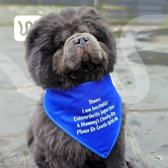
Thanos, a three-year-old Chow Chow, had bilateral tarsal hyperextension alongside hip and elbow dysplasia and spinal disease. His clinical team selected a WIMBA Tarsus Orthosis GO with motion-control Tokens to support the hock during prescribed activity. The published observations describe one complex patient and do not predict the outcome of another dog.14
Provider: Kate Outram Veterinary Physiotherapy
Solution: WIMBA Tarsus Orthosis GO
Fitting, Skin Checks, and Follow-Up
Managing tarsal instability in dogs with any custom dog tarsal brace requires professional fitting and reassessment. The first fitting should take place with the WIMBA Provider, who checks joint-axis alignment, frame position, strap tension, insert placement around the Achilles region, rotation, migration, and the dog’s gait.13
- Follow the prescribed acclimatization plan. There is no universal wear schedule for every form of tarsal instability in dogs.
- Supervise device use. WIMBA orthoses are not intended for continuous 24-hour wear or unattended activity.
- Inspect the skin after use. Check for persistent redness, hair loss, rubbing, swelling, abrasions, heat, or moisture.
- Watch the device during walking. It should not rotate, slide, contact the limb with hard frame elements, or change the gait in a concerning way.
- Keep the limb and device clean and dry. Follow the product care instructions provided by the clinic.
- Return for review. Changes in swelling, muscle mass, body weight, coat, activity, pain, or disease progression can alter fit and the required motion settings.
Remove the orthosis and contact the provider if persistent redness, a sore, new swelling, new pain, repeated migration, device damage, or worsening lameness develops. Do not cut, heat, drill, bend, or reshape the frame at home. Safe management of tarsal instability in dogs depends on the clinical plan continuing after delivery.
Questions to Ask the Veterinary Team
Before choosing external support for tarsal instability in dogs, make sure the clinical objective and follow-up plan are clear.
- Which structure and joint level are causing the abnormal hock position?
- Is the problem hyperextension, hyperflexion, side-to-side instability, or a functional gait pattern?
- Does the tendon, ligament, fracture, or joint damage require surgery?
- If external support is appropriate, what movement must the device restrict or allow?
- Would GO or PRO match the plane and severity of this dog hock instability?
- What activity, wear, skin-check, and rehabilitation schedule should we follow?
- Which findings mean the device should be removed and the dog rechecked?
- How will progress be measured: gait, pain, function, imaging, range of motion, or another outcome?
Frequently Asked Questions
What is tarsal instability in dogs?
Tarsal instability in dogs is abnormal control or motion at the hock region. It may involve excessive extension, excessive flexion, side-to-side deviation, rotation, or subluxation caused by ligament, tendon, bone, joint, neurological, or compensatory problems. It is a descriptive finding, so the affected structure still has to be diagnosed.
Does a dropped hock mean tarsal hyperextension or an Achilles injury?
Either may be described as a dropped hock, but the mechanics differ. Failure of plantar supporting structures can produce excessive tarsal extension, while common calcaneal tendon injury usually allows excessive tarsal flexion and may create a plantigrade stance. Examination and imaging are needed to distinguish them.
Can tarsal instability in dogs be treated without surgery?
Selected mild, partial, stable, or chronic cases may be managed without surgery using veterinarian-directed activity control, rehabilitation, pain management, and external support. Complete tendon rupture, severe ligament failure, fracture-luxation, or major multiplanar instability may require surgical repair or arthrodesis.
What are the non-surgical treatment options for tarsal (ankle) instability in dogs?
Once a veterinary assessment confirms that conservative management is appropriate, the plan may combine prescribed activity restriction, pain management, weight management where relevant, and joint-specific rehabilitation introduced at the correct stage of healing. External motion control may also be included: WIMBA Tarsus Orthosis GO is intended for selected mild, predominantly single-plane cases, while a PRO pathway may be considered when more advanced or multiplanar control is required. A brace supports the joint but does not heal a complete tendon rupture or replace surgical repair or arthrodesis when either is indicated.
When should a dog with suspected hock instability see a veterinarian?
Arrange an examination when the hock repeatedly collapses, angles abnormally, swells, becomes painful, or changes the dog’s gait. Seek prompt care after major trauma or if the dog cannot bear weight, has an open wound or obvious deformity, develops rapidly increasing swelling, or shows worsening weakness or poor coordination.
Can a dog tarsal brace replace surgery?
No. A dog tarsal brace can provide external motion control in a suitable, diagnosed case, but it does not repair a complete tendon rupture, reconstruct a severely failed ligament, or stabilize every fracture or luxation. It may complement conservative care or postoperative rehabilitation when the treating veterinary team defines that role.
How is a WIMBA Tarsus Orthosis fitted?
For eligible GO cases, the clinic submits two required limb photos and product-specific manual measurements through WimbaAPP. PRO uses WimbaSCAN plus manual measurements for more complex cases. The finished orthosis is delivered to the clinic for professional fitting, gait assessment, owner instruction, and follow-up.
What does veterinary rehabilitation involve for dogs with tarsal instability?
When a veterinarian determines that conservative management or postoperative recovery is appropriate, a rehabilitation professional may introduce controlled exercises, weight-bearing retraining, and joint-specific work at the correct stage of healing — never before the diagnosed structure has been assessed. Rehabilitation does not replace surgical repair when a complete tendon rupture, severe ligament failure, or unstable fracture requires it, but it can support recovery of strength, coordination, and function alongside activity control, pain management, and external bracing when the veterinary team includes it in the plan.
Start With the Cause of the Hock Instability
The same visible posture can come from very different injuries. If your dog shows possible tarsal instability, begin with a veterinary diagnosis. Pet owners can find an experienced WIMBA Provider, while veterinary professionals can submit the clinical details for case review.
Medical disclaimer: This article is intended for general educational purposes only and is not a substitute for veterinary examination, diagnosis, or treatment. Do not manipulate an unstable joint, apply a rigid device, start rehabilitation exercises, or change prescribed activity without professional direction. Seek prompt veterinary care after significant trauma or if your dog cannot bear weight, has an open injury or obvious deformity, shows severe pain, or develops progressive weakness or poor coordination.
Scientific, Clinical, and Product References
- Harasen G. Arthrodesis—Part II: The tarsus. Canadian Veterinary Journal. 2002;43(10):806–808. Full text.
- Abako J, Holak P, Głodek J, Zhalniarovich Y. Usefulness of Imaging Techniques in the Diagnosis of Selected Injuries and Lesions of the Canine Tarsus: A Review. Animals. 2021;11(6):1834. doi:10.3390/ani11061834.
- Gamble LJ, Canapp DA, Canapp SO. Evaluation of Achilles Tendon Injuries With Findings From Diagnostic Musculoskeletal Ultrasound in Canines—43 Cases. Veterinary Evidence. 2017;2(3). doi:10.18849/ve.v2i3.92.
- American College of Veterinary Surgeons. Achilles’ Tendon Injuries. Clinical resource.
- Jung W, Kim HM, Son SJ, Kim HY. Bilateral Functional Tarsal Hyperextension Associated with Stifle Pathology Improved Following Stifle Stabilization in a Dog: A Case Report. Veterinary Sciences. 2026;13(6):518. doi:10.3390/vetsci13060518.
- Wylie S, Piana F, Montgomery P, et al. Complications and outcomes following surgical management of common calcaneal tendon pathology in 80 dogs. Veterinary Surgery. 2026;55(2):437–447. doi:10.1111/vsu.70053.
- Lister SA, Renberg WC, Roush JK. Efficacy of immobilization of the tarsal joint to alleviate strain on the common calcaneal tendon in dogs. American Journal of Veterinary Research. 2009;70(1):134–140. doi:10.2460/ajvr.70.1.134.
- Case JB, Palmer R, Valdes-Martinez A, Egger EL, Haussler KK. Gastrocnemius tendon strain in a dog treated with autologous mesenchymal stem cells and a custom orthosis. Veterinary Surgery. 2013;42(4):355–360. doi:10.1111/j.1532-950X.2013.12007.x.
- Rosen S, Duerr FM, Elam LH. Prospective evaluation of complications associated with orthosis and prosthesis use in canine patients. Frontiers in Veterinary Science. 2022;9:892662. doi:10.3389/fvets.2022.892662.
- WIMBA. WIMBA Tarsal Orthotics GO. Product and ordering information.
- WIMBA Knowledge Base. WIMBA GO Orthotics Indications. Current clinical indications.
- WIMBA. WIMBA Orthotics PRO. Product and ordering information.
- WIMBA Knowledge Base. WIMBA Tarsus Orthosis GO—First Donning. Fitting guidance.
- WIMBA. Tarsal Hyperextension in Dogs: Case Study Thanos. Individual observational case report.
- WIMBA Knowledge Base. WIMBA Orthosis Delivery Times. Current production information.
Related reading: Achilles Tendon Injury in Dogs: Signs, Treatment, and Orthotic Support. · Custom Dog Hock Orthotics for Daily Walking Support