 Skip to content

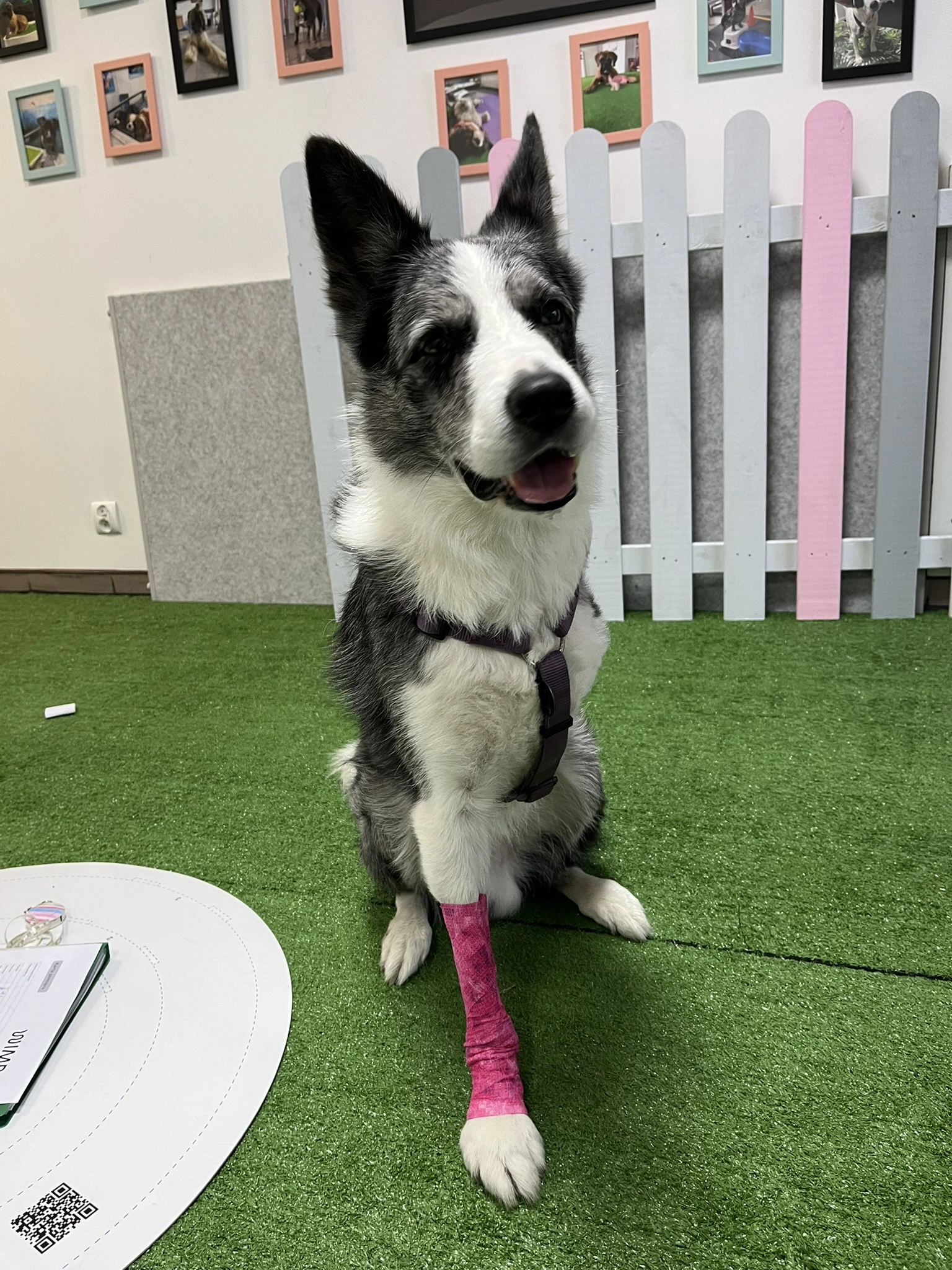
If your dog has just become, or is about to become, a tripod, this is the starting point. Three-legged dog mobility support covers the basics every new owner and referring clinic needs first: how limb loss changes gait and weight distribution, what to expect week by week, and when to move on to more specific questions about orthotics, wheelchairs, or rehabilitation covered elsewhere on this site. Many dogs adapt impressively after amputation, but successful adaptation does not remove the need for long-term monitoring and a structured mobility plan.
Effective tripod dog support may combine veterinary assessment, rehabilitation, weight management, home modification, and targeted orthotic intervention. This guide explains the evidence behind post-amputation mobility and shows when a custom device may support a clearly identified joint or movement problem.
Looking for something more specific? This guide focuses on veterinary assessment, candidate selection, and the fitting process for tripod dogs. For WIMBA patient stories and the current ordering process, see Tripod Dog Orthotics and Mobility Support. For a joint-by-joint guide and a clear explanation of how orthoses differ from prostheses, see Orthotics for Three-Legged Dogs.
Why Three-Legged Dog Mobility Support Matters
Most owners report favourable adaptation after canine limb amputation. In a retrospective study of 64 dogs, 88% of owners reported a complete or nearly complete return to their dog’s pre-amputation quality of life, and 78% felt that recovery and adaptation were better than expected.[1] These encouraging owner-reported outcomes show what tripod dogs can achieve, while also highlighting the importance of body weight and body condition, which were negatively associated with quality-of-life scores in the same study.
Adaptation requires a new movement strategy. Studies using force platforms, stance analysis, electromyography, and pressure-sensitive walkways consistently show changes in limb loading, stance time, spinal motion, and muscle recruitment after the loss of a limb.[2][3][4] A proactive plan for three-legged dog mobility support uses those findings to guide monitoring without assuming that every compensatory change will become a clinical problem.
What current gait research shows:
- Dogs redistribute load across all three remaining limbs after amputation
- The remaining front limb often carries the largest increase in standing load
- Front-limb and hind-limb amputees use different compensatory strategies
- Spinal motion and muscle recruitment change as the dog establishes a tripedal gait
- Clinical decisions should be based on the individual dog’s examination, gait, comfort, and functional goals
How Limb Loss Changes Gait and Loading
After Front-Limb Amputation
The remaining front limb becomes especially important after front-limb amputation. In a study comparing 16 front-limb amputees with 24 four-limbed control dogs at a trot, weight distribution increased by 14% on the remaining front limb and by a combined 17% on the hind limbs. The amputee dogs also showed changes in stance duration, braking forces, joint position, and spinal movement.[3]
A 2024 pressure-walkway study involving 39 amputee dogs reported that the contralateral front limb carried 50.7% of body weight after high front-limb amputation and 55.5% after low front-limb amputation during the measured walking trials.[6] These figures describe the studied groups and should not be treated as a prediction for every dog, but they explain why the carpus and the rest of the remaining front limb deserve regular assessment.
After Hind-Limb Amputation
Hind-limb amputees redistribute braking and propulsive work across the remaining hind limb and both front limbs. A kinetic and kinematic study found increased braking forces in the opposite front limb, increased propulsive forces and impulses in the front limb on the amputated side and the remaining hind limb, and altered movement at the tarsus and several spinal regions.[4]
For long-term dog mobility after amputation, this means that examination should extend beyond the limb opposite the amputation. The front limbs, remaining hind limb, pelvis, and spine all contribute to the dog’s new movement strategy and may require different rehabilitation priorities.
Trunk and Muscle Adaptation
Tripod locomotion also changes how the dog recruits limb and back muscles. In an experimental study that simulated loss of hind-limb function in nine dogs, researchers observed changes in the timing and intensity of muscle activity in the remaining hind limb, front limb, and long back muscle.[5] The study demonstrated coordinated muscular adaptation, but it did not establish that specific exercises or devices prevent future spinal disease.
What Orthotic Support for Tripod Dogs Can Do
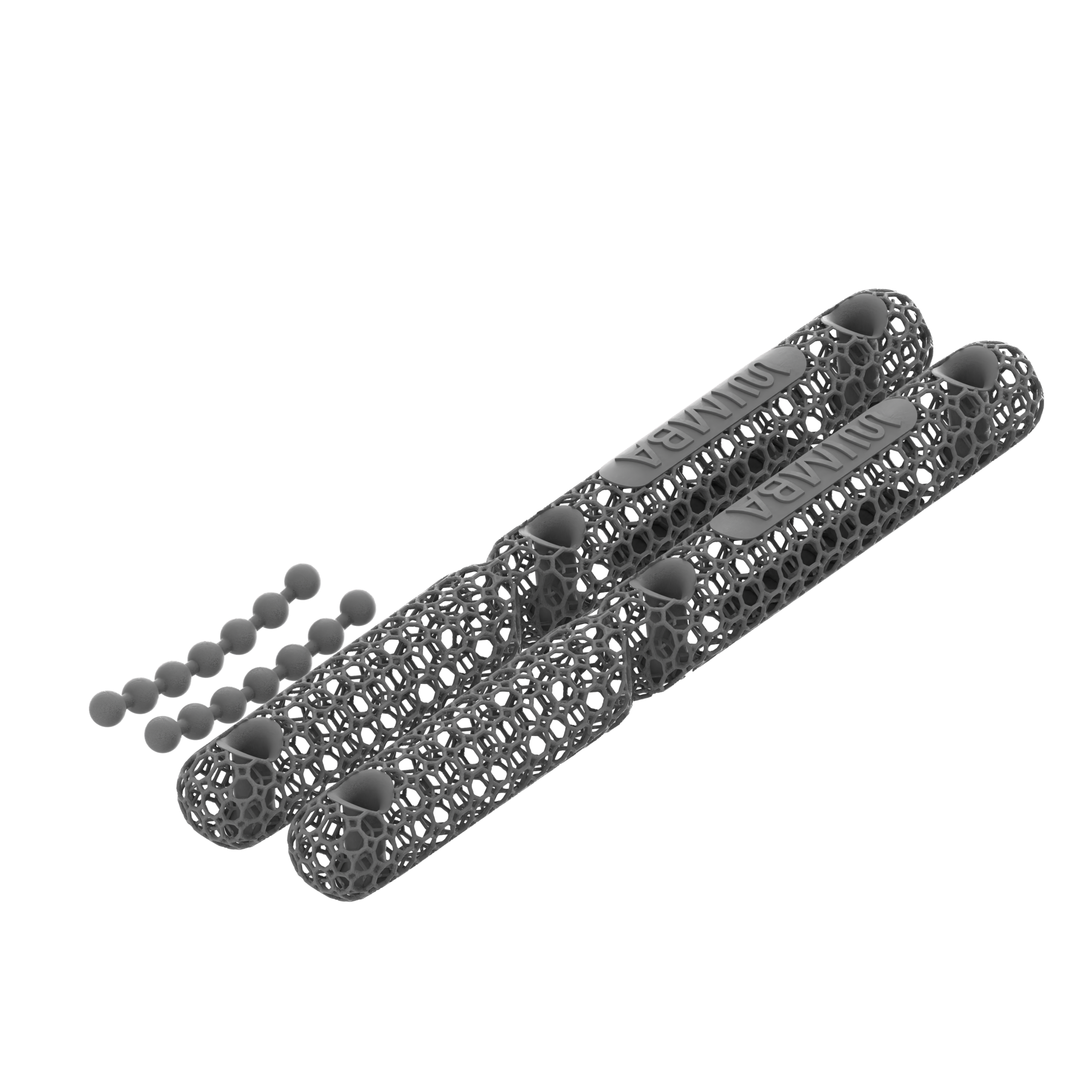
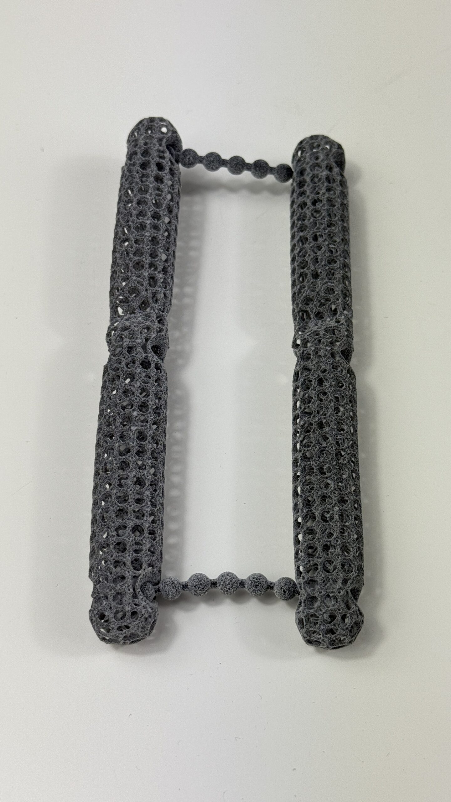
Orthotic support for tripod dogs is directed at an existing limb or joint. An orthosis can be designed to restrict a selected movement, guide alignment, support an unstable joint, or protect tissue during controlled activity. A prosthesis serves a different purpose by replacing a missing limb or limb segment. This article and WIMBA’s tripod pathway focus on orthoses for the remaining limbs.
A dog orthosis after amputation should be prescribed for a defined mechanical target. Examples include documented carpal hyperextension, tarsal instability, a clinician-selected range-of-motion goal, or support during a rehabilitation protocol. The device does not reverse limb loss, eliminate all compensatory loading, or guarantee prevention of osteoarthritis or ligament injury.
A clinically selected orthosis may help to:
- Limit repeated carpal or tarsal hyperextension
- Support a diagnosed single-plane or multiplanar instability
- Control range of motion during a veterinary rehabilitation plan
- Support clinician-selected joint alignment during loading
- Provide a repeatable external support strategy during selected activities
The strongest tripod dog support plans connect the device to an examination finding and a measurable goal. Early orthotic assessment helps the veterinary team identify where controlled joint support may add value, while ensuring that imaging, analgesia, surgery, or another indicated treatment is incorporated into the wider plan.
When to Consider a Dog Orthosis After Amputation
Carpal Hyperextension or Laxity
The carpus of the remaining front limb is a priority for assessment, especially after front-limb amputation. Visible sinking through the wrist, increasing extension during stance, reduced endurance, or a change in paw placement should prompt veterinary examination. A carpal orthosis may be used to limit excessive extension when the clinician confirms that this is the relevant mechanical problem.
Tarsal or Stifle Instability
After hind-limb amputation, the remaining tarsus and stifle should be monitored alongside both front limbs. A tarsal or stifle orthosis may be considered when examination and imaging identify instability, an injury, osteoarthritis affecting the supported joint, or a postoperative goal. Bracing should not be presented as an automatic substitute for surgical stabilisation.
Changes in Gait, Endurance, or Comfort
Reduced walking tolerance, repeated stopping, altered posture, reluctance on stairs, slipping, joint swelling, or a new change in gait deserves assessment rather than being attributed automatically to age. A dog orthosis after amputation may form part of the response when the examination identifies a joint that can benefit from external control.
Assessment and Candidate Selection
A useful prescription starts with a whole-patient assessment. The veterinary team should establish which structure requires support, which movement should be controlled, and how success will be measured. This prevents three-legged dog mobility support from becoming a generic brace recommendation without a clinical objective.
Before selecting a device, assess:
- The reason for amputation, healing status, pain, and current medication
- Standing posture, gait, endurance, and movement on relevant surfaces
- All remaining limbs, not only the limb opposite the amputation
- Joint range of motion, instability, swelling, and muscle condition
- Skin, coat, sensation, wounds, and pressure-sensitive areas
- Body condition, home environment, activity level, and owner goals
- The owner’s ability to fit, remove, clean, and inspect the device
Baseline photographs, video, measurements, and functional observations make follow-up more useful. They allow the team to distinguish genuine improvement from day-to-day variation and to identify when the prescription, rehabilitation plan, or device settings need review.
Fitting, Break-In, and Follow-Up
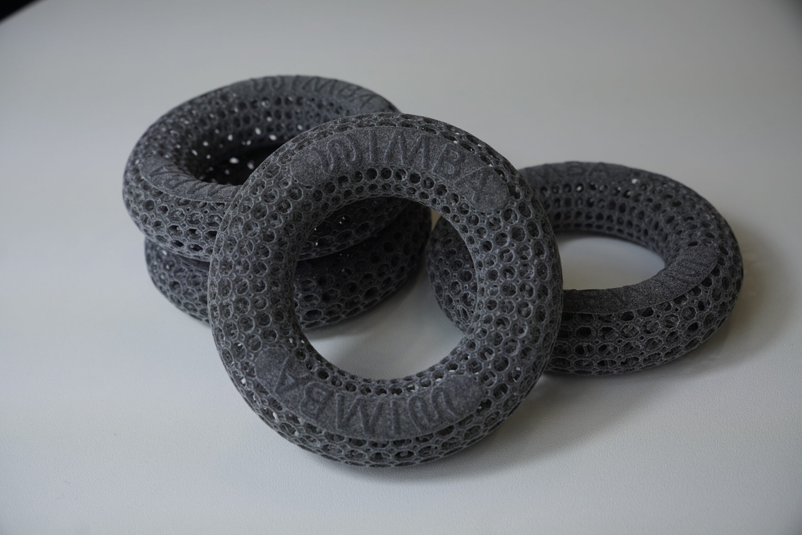
Custom fabrication can accommodate the anatomy of the remaining limb and the selected mechanical objective. Digital scanning can capture external limb geometry without plaster casting, while guided photographs and manual measurements may be suitable for less complex cases. The production method alone does not guarantee a good outcome. Prescription, fit, device design, introduction, and follow-up all matter.
Every dog orthosis after amputation requires a gradual break-in schedule set by the prescribing veterinary professional. Owners should check the skin after each wearing period and report persistent redness, swelling, rubbing, pain, device migration, or worsening gait. The device should be removed and reviewed if any of these problems occur.
Complications are a practical reason for planned follow-up. In a prospective study of 43 dogs using carpal, tarsal, or stifle orthoses or prostheses, at least one complication was recorded in 39 dogs. Skin complications were particularly common during the first three months.[7] The study included different device types and conditions, so its results should not be interpreted as a device-specific complication rate for tripod dogs.
Rehabilitation and Dog Mobility After Amputation
Rehabilitation and orthotic management serve different but complementary functions. An orthosis can control a selected movement externally, while rehabilitation develops strength, coordination, endurance, confidence, and safe movement strategies. A coordinated plan for dog mobility after amputation may include controlled walking, progressive strengthening, balance work, range-of-motion exercises, and hydrotherapy when the surgical site and clinical condition allow it.
Exercise selection should be individual. The location of amputation, concurrent osteoarthritis, neurological status, cancer treatment, cardiopulmonary fitness, age, and previous activity all influence what is appropriate. Swimming, underwater treadmill work, cavaletti exercises, and balance equipment should be introduced by the rehabilitation team rather than copied from a generic online programme.
Weight management is a central part of tripod dog support. In the owner-outcome study described earlier, greater body weight and body condition at amputation were negatively correlated with reported quality of life.[1] Maintaining an appropriate body condition reduces avoidable demand on the remaining limbs and makes controlled exercise easier.
The WIMBA Pathway for Tripod Patients
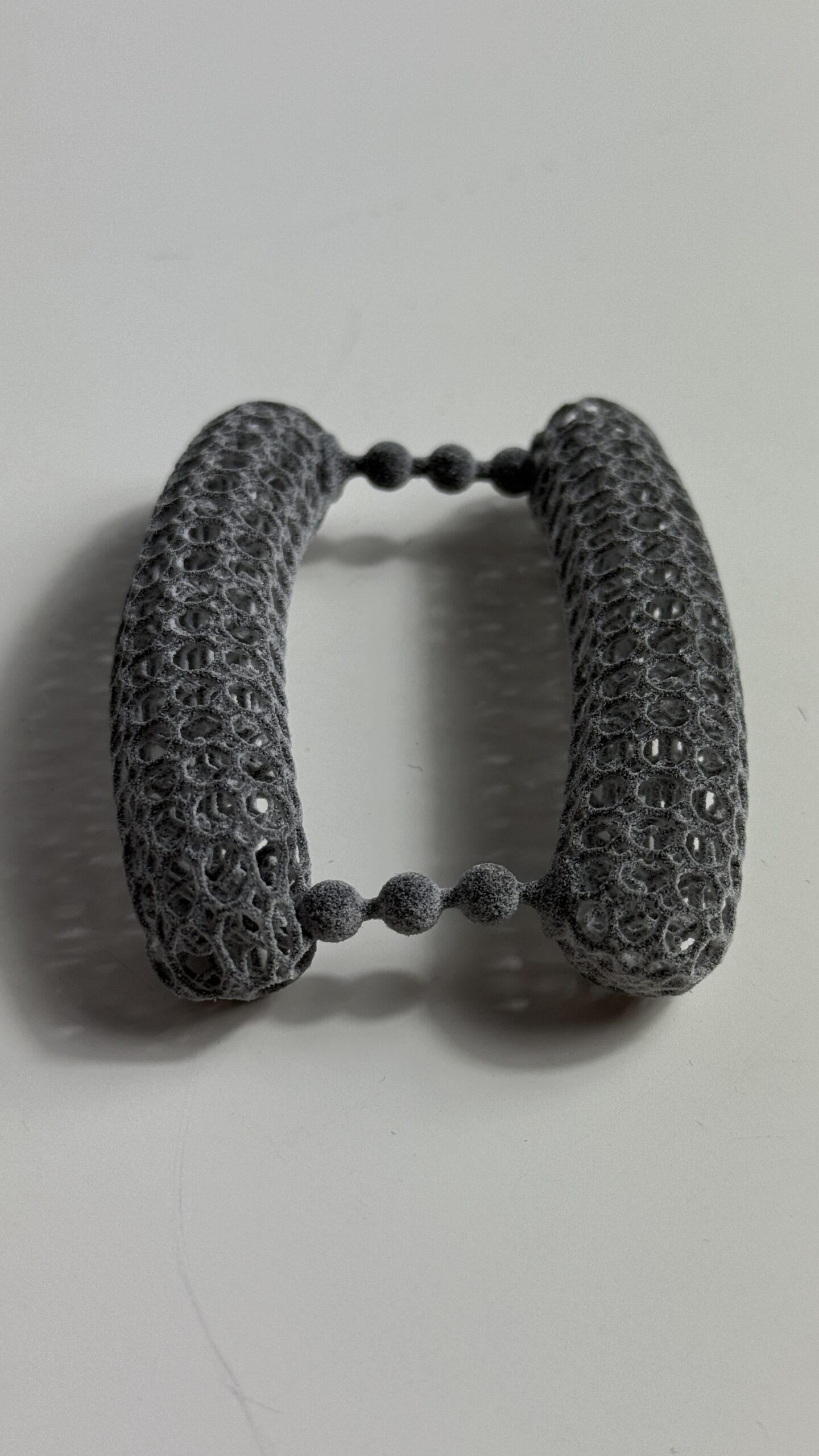
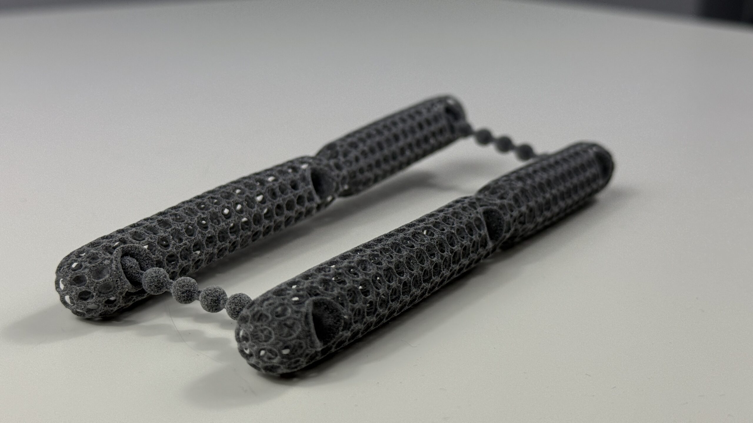
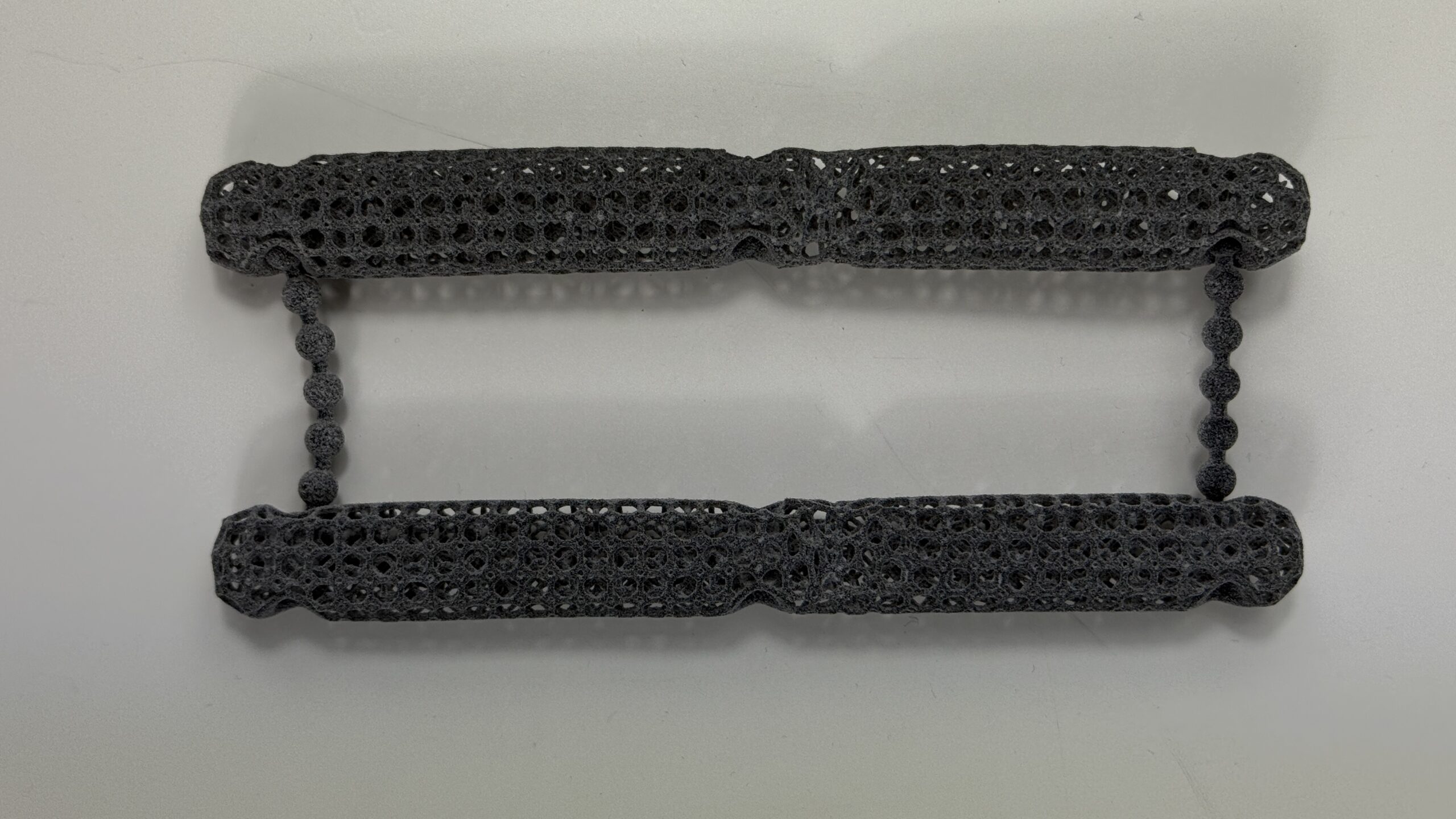
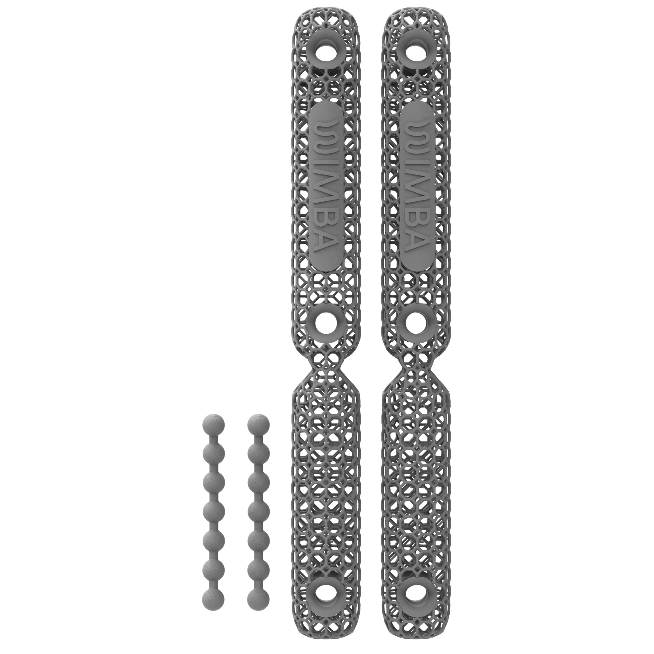
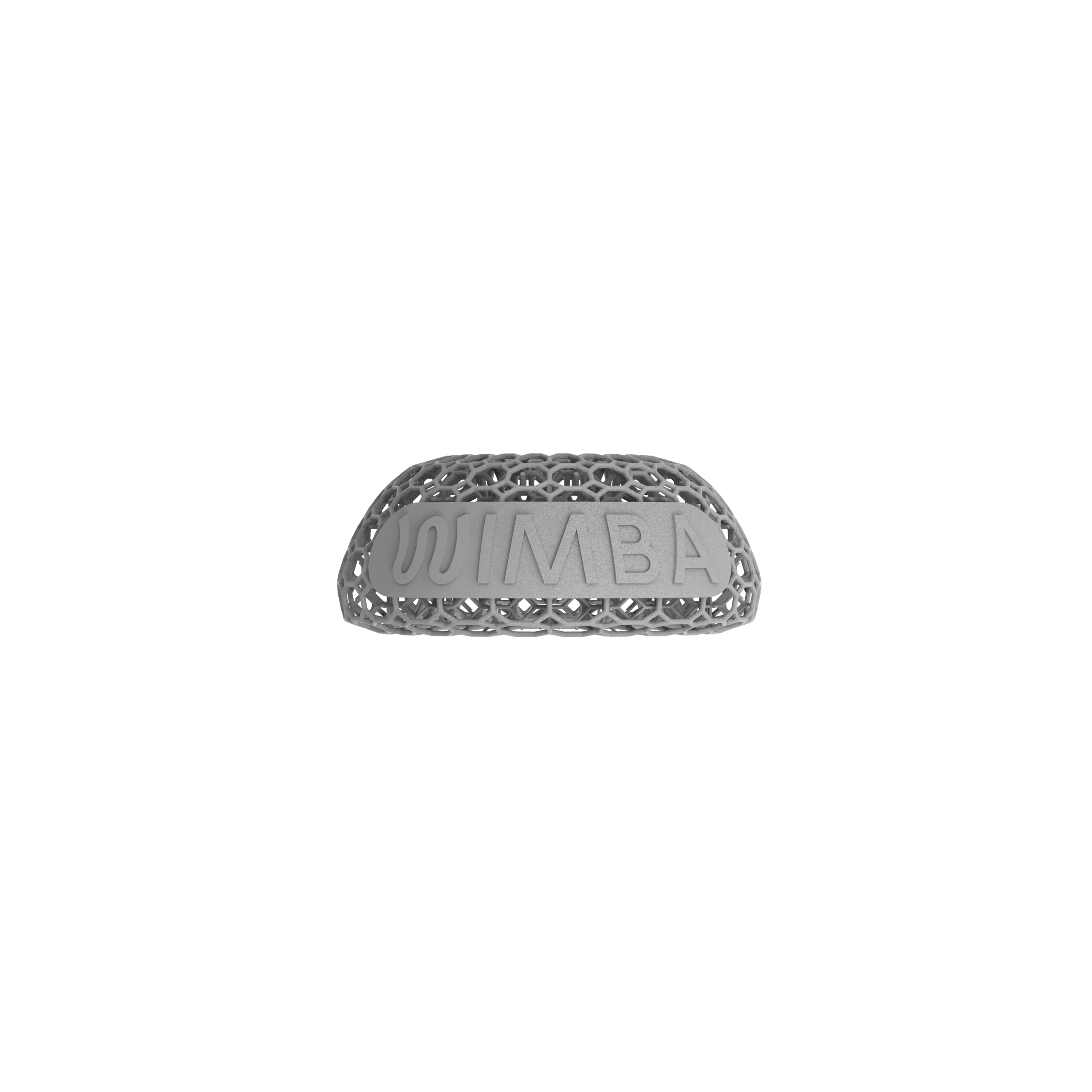
WIMBA provides custom, 3D-printed orthoses for existing limbs. Within a three-legged dog mobility support plan, the aim is to match the device to the diagnosed joint problem and the veterinary team’s functional goal rather than to amputation status alone.
WIMBA GO uses guided photographs and manual measurements for mild, less complex carpal and tarsal cases. WIMBA PRO uses WimbaSCAN for patient-specific carpal, tarsal, and stifle devices when more complex or multiplanar control is required. Both pathways use HP Multi Jet Fusion manufacturing and should be integrated with a clinician-defined wearing and follow-up plan.
Veterinary professionals can request a no-cost case consultation to discuss orthotic support for tripod dogs, device selection, and the appropriate GO or PRO pathway. Pet owners can use the WIMBA Provider Map to find a participating veterinary or rehabilitation clinic. WIMBA works through veterinary professionals, who remain responsible for diagnosis, prescription, fitting, and follow-up.
For a practical example, read Juri’s case study, which describes how a WIMBA Carpus Orthosis GO was incorporated after front-limb amputation when the remaining carpus already showed degenerative change and a tendency towards hyperextension.
Frequently Asked Questions: Three-Legged Dog Mobility Support
Can a three-legged dog live an active life?
Many tripod dogs return to walking, play, and enjoyable daily activity after amputation. In one retrospective study, 88% of owners reported complete or nearly complete return to pre-amputation quality of life. A structured three-legged dog mobility support plan helps the veterinary team coordinate weight management, rehabilitation, pain assessment, and monitoring as the dog’s needs change.
Why should the remaining front limb be monitored?
Studies of standing and walking show that the remaining front limb often takes the largest increase in load after amputation. This is especially relevant after front-limb loss, when the opposite carpus becomes the only front wrist accepting ground contact. Monitoring can identify changes in posture, hyperextension, comfort, and endurance before they become more difficult to manage.
What should I do in the first few weeks after my dog becomes a tripod?
Focus on a safe recovery environment, following the surgical team’s activity restrictions, and a scheduled recheck, rather than any single device decision. Orthotic or mobility-aid questions come later, once the surgical site has healed and a clinician can properly assess gait and loading; this guide gives a starting checklist for that early period before pointing you to more specific resources.
What orthotic support for tripod dogs is available?
Orthotic support for tripod dogs may include a carpal, tarsal, or stifle device, depending on the affected joint and mechanical objective. Front-limb amputees are often assessed for carpal hyperextension in the remaining front limb. Hind-limb amputees may require assessment of the remaining tarsus or stifle, as well as both front limbs.
How can owners support dog mobility after amputation?
Owners can maintain an appropriate body condition, follow the rehabilitation plan, use non-slip surfaces, monitor gait and skin, and report changes promptly. Effective dog mobility after amputation also depends on regular veterinary reassessment rather than waiting for severe lameness or loss of endurance.
How does a custom orthosis fit into tripod dog support?
Within tripod dog support, a custom orthosis addresses a defined problem in an existing joint. It may limit excessive movement, guide alignment, or support controlled rehabilitation. It works alongside veterinary care and exercise rather than replacing diagnosis, pain management, or surgery when another treatment is indicated.
How can a clinic request WIMBA support?
Veterinary professionals can submit the patient’s history, diagnosis, photographs, gait information, and intended functional goal through WIMBA’s case consultation form. The WIMBA clinical team can help identify the appropriate GO or PRO pathway, while the treating veterinary professional retains responsibility for clinical decisions and follow-up.
What should a new tripod dog owner know about day-to-day care?
Day-to-day care should follow the veterinary team’s recovery and activity plan. Maintain a lean body condition, increase exercise gradually as directed, provide secure non-slip footing, and monitor the surgical site during healing as well as gait, comfort, fatigue, swelling, and the remaining limbs. New lameness, reduced activity, swelling, skin changes, or worsening exercise tolerance should prompt veterinary review. An orthosis should be considered only when examination identifies a specific joint or mechanical problem; tripod status alone is not an indication for routine bracing.
Medical Disclaimer: All information on this website is intended for instruction and information purposes only. The authors are not responsible for any harm or injury that may result. Significant injury risk is possible if you do not seek suitable professional advice about your patient’s specific situation. No guarantees of specific results are expressly made or implied on this website.
Scientific References
- Dickerson VM, Coleman KD, Ogawa M, et al. Outcomes of dogs undergoing limb amputation, owner satisfaction with limb amputation procedures, and owner perceptions regarding postsurgical adaptation: 64 cases (2005–2012). J Am Vet Med Assoc. 2015;247(7):786–792. doi:10.2460/javma.247.7.786
- Cole GL, Millis D. The effect of limb amputation on standing weight distribution in the remaining three limbs in dogs. Vet Comp Orthop Traumatol. 2017;30(1):59–61. doi:10.3415/VCOT-16-05-0075
- Jarvis SL, Worley DR, Hogy SM, Hill AE, Haussler KK, Reiser RF II. Kinematic and kinetic analysis of dogs during trotting after amputation of a thoracic limb. Am J Vet Res. 2013;74(9):1155–1163. doi:10.2460/ajvr.74.9.1155
- Hogy SM, Worley DR, Jarvis SL, Hill AE, Reiser RF II, Haussler KK. Kinematic and kinetic analysis of dogs during trotting after amputation of a pelvic limb. Am J Vet Res. 2013;74(9):1164–1171. doi:10.2460/ajvr.74.9.1164
- Fuchs A, Anders A, Nolte I, Schilling N. Limb and back muscle activity adaptations to tripedal locomotion in dogs. J Exp Zool A Ecol Genet Physiol. 2015;323(8):506–515. doi:10.1002/jez.1936
- Filho TG, Rahal SC, Kano WT, et al. Gait analysis of amputee dogs using a pressure-sensitive walkway. Vet Comp Orthop Traumatol. 2024;37(4):189–195. doi:10.1055/s-0044-1779727
- Rosen S, Duerr FM, Elam LH. Prospective evaluation of complications associated with orthosis and prosthesis use in canine patients. Front Vet Sci. 2022;9:892662. PMC9372342