 Skip to content


A veterinary orthosis can support selected neurological, orthopaedic, postoperative, and compensatory mobility problems in dogs and cats. The right device may restrict excessive joint motion, guide alignment, protect vulnerable tissues, or assist controlled weight-bearing during rehabilitation.
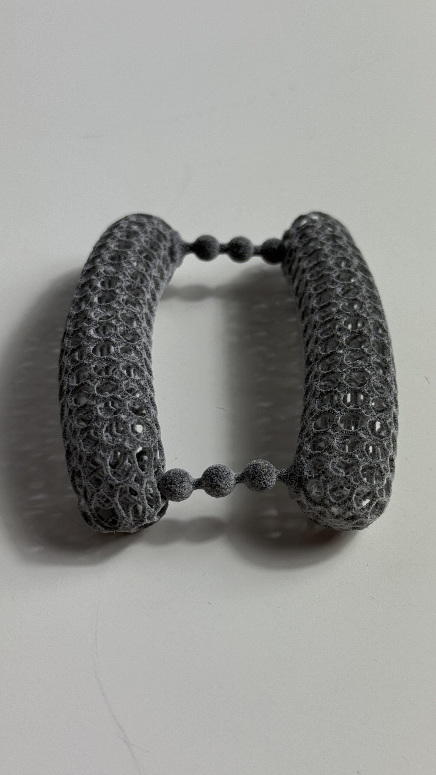
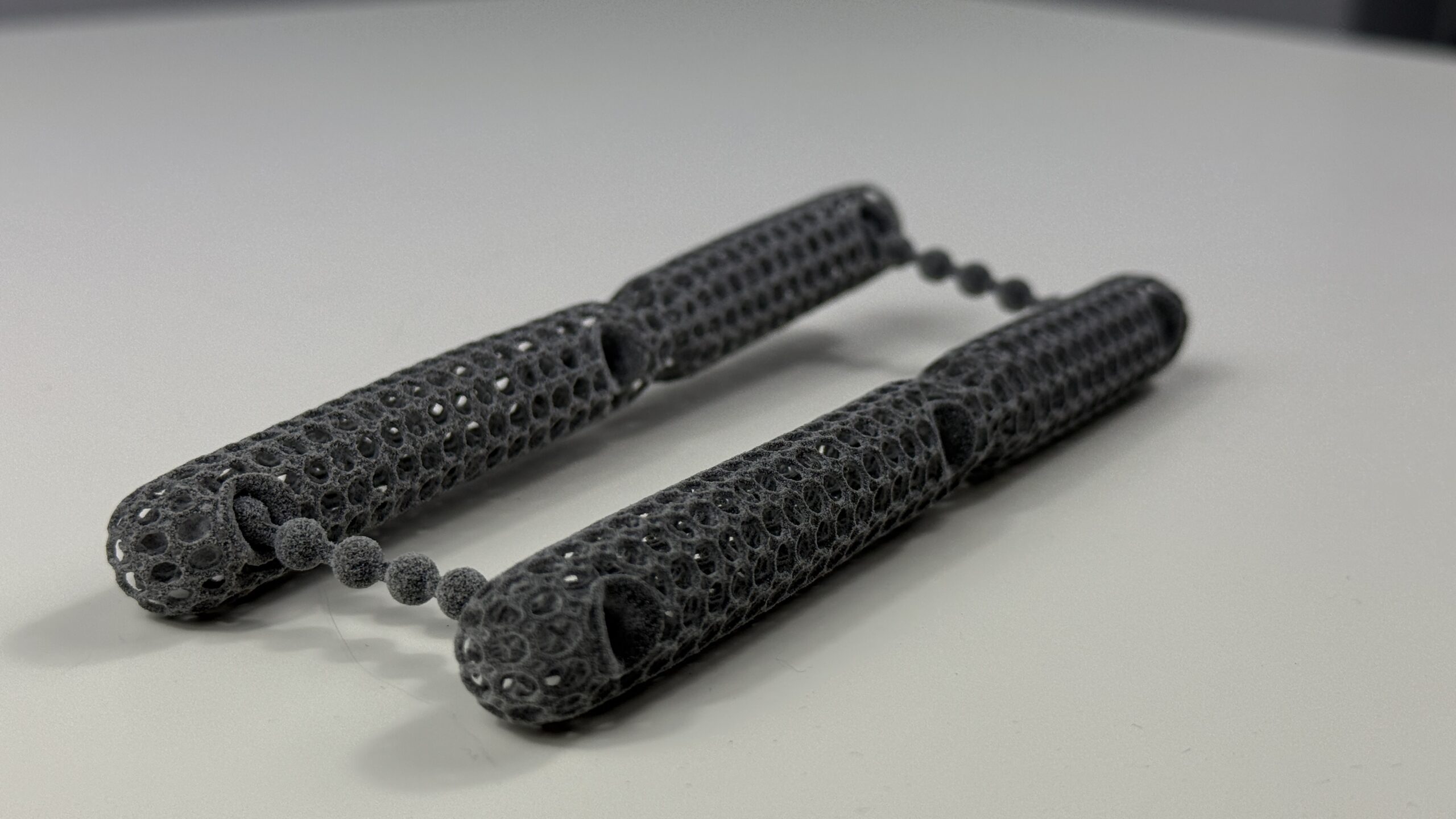
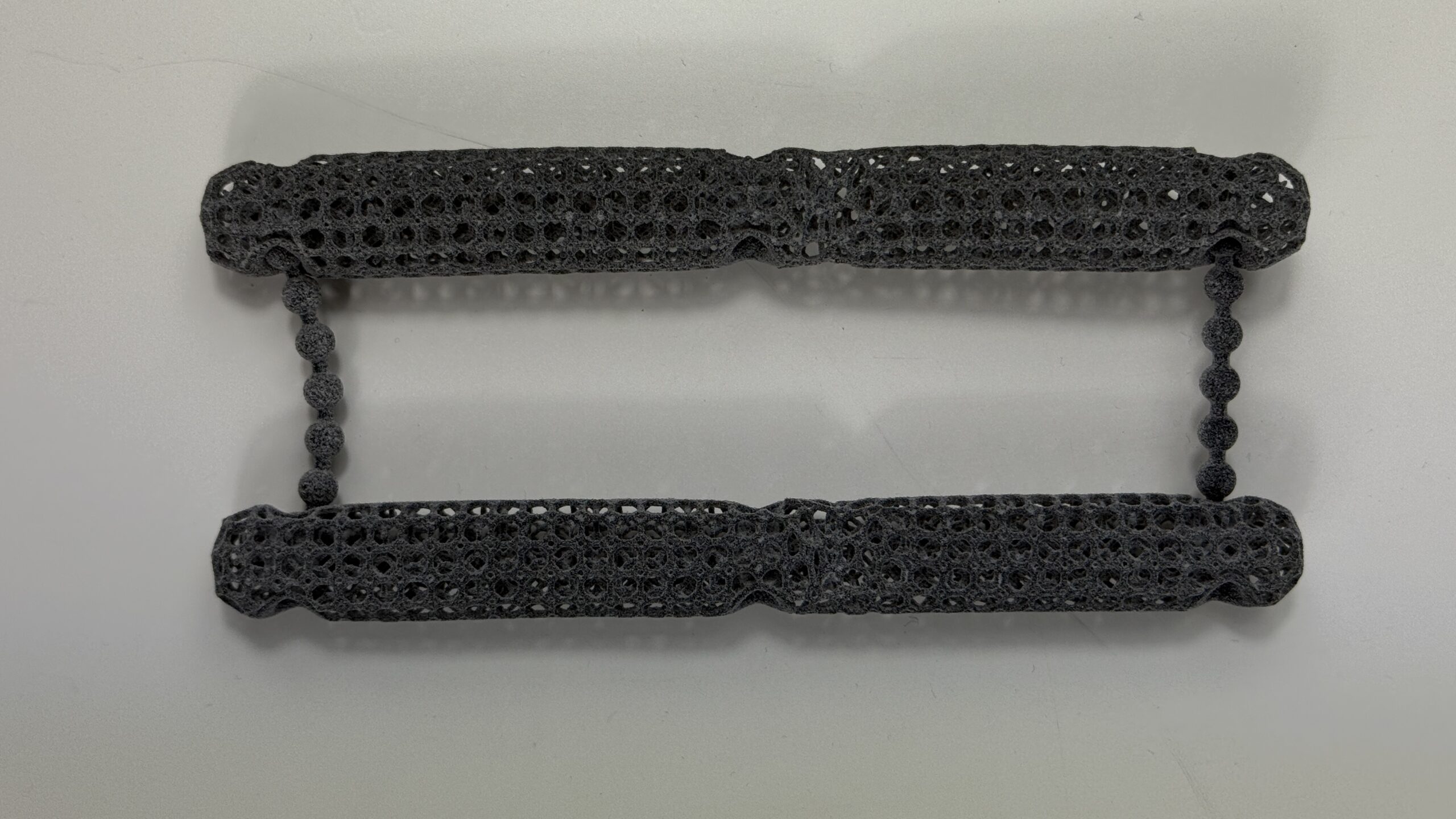
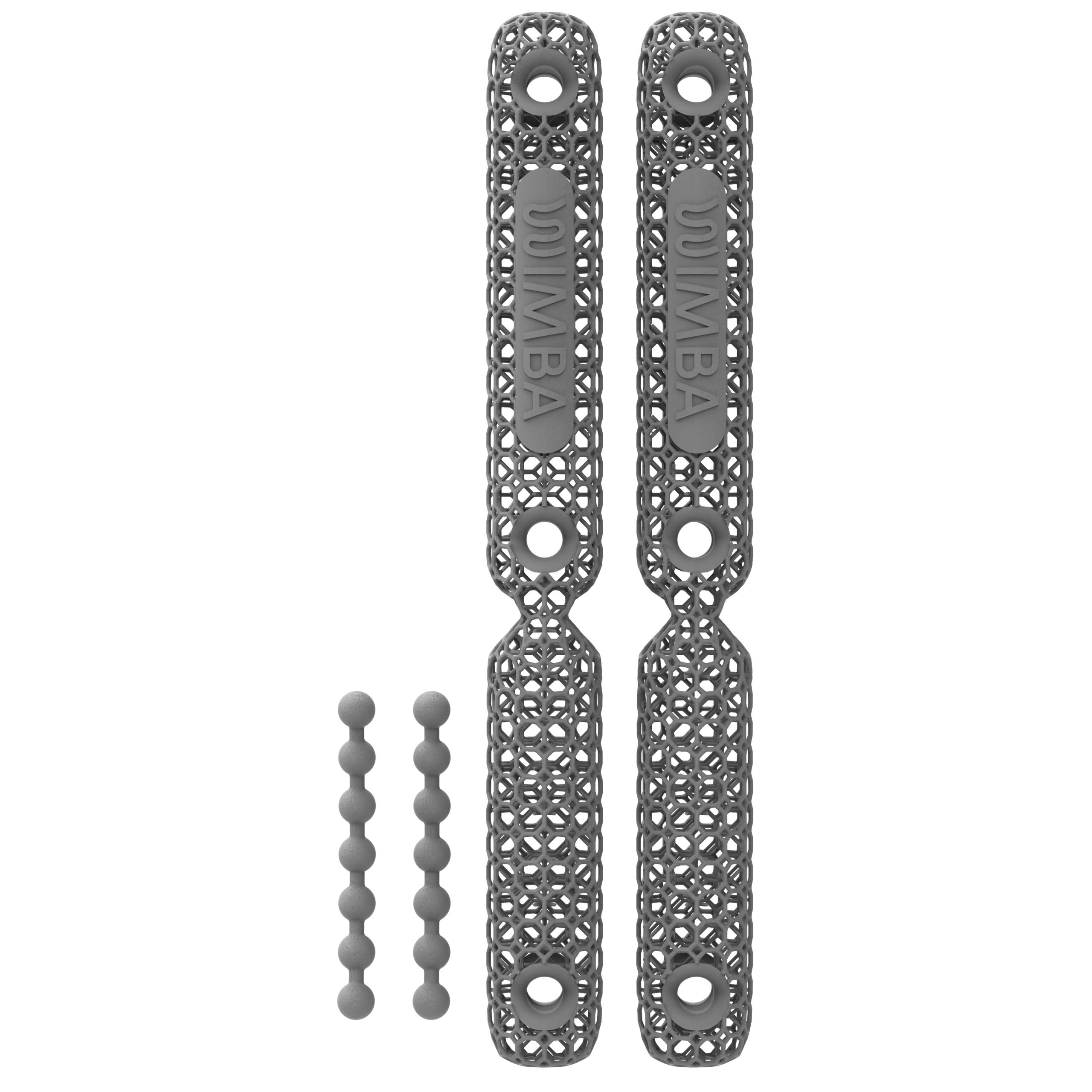
WIMBA makes custom orthoses for dogs and cats, including carpal, tarsal and stifle devices, used under veterinary guidance to support selected orthopaedic, neurological and postoperative cases.
This guide explains the main clinical indications for custom dog orthotics, where the evidence remains limited, and how veterinary teams can identify suitable candidates without presenting dog braces as substitutes for diagnosis, rehabilitation, or surgery when surgery is indicated.
What Veterinary Orthoses Can and Cannot Do
An orthosis is an external medical device that supports an existing joint or limb segment. Its design should begin with a defined mechanical objective: which movement should be allowed, which movement should be restricted, and what functional task should become safer or more comfortable for the patient?
Depending on the diagnosis and design, an orthosis may help to:
- Limit excessive flexion, extension, rotation, or side-to-side instability
- Maintain a clinician-selected joint position or controlled range of motion
- Support a weakened carpal, tarsal, or stifle joint during daily activity
- Protect the paw from abrasion associated with toe dragging or altered placement
- Assist controlled loading within a veterinary rehabilitation plan
A veterinary orthosis does not establish a diagnosis, decompress the spinal cord or nerve roots, repair a completely ruptured ligament or tendon, or reverse a progressive neurological disease. It should not delay surgical referral when instability, pain, or neurological deficits make surgery the recommended treatment. Dog orthotics are one component of a wider plan that may also include medication, weight management, therapeutic exercise, physiotherapy, hydrotherapy, and environmental modification.
Neurological Conditions and Dog Orthotics
Degenerative Myelopathy (DM)
Degenerative myelopathy is a progressive, typically non-painful spinal cord disease associated with proprioceptive ataxia and paresis of the pelvic limbs. Rehabilitation remains central to maintaining strength, coordination, and function. In a retrospective study of dogs with suspected degenerative myelopathy, intensive controlled physiotherapy was associated with longer survival than moderate or no physiotherapy, but this study did not evaluate orthoses.[1]
In neurological cases, canine orthotics may be considered for an ambulatory patient when there is a specific mechanical goal, such as protecting the dorsal paw from abrasion, improving toe clearance, or supporting documented carpal or tarsal instability. An orthosis does not slow or reverse the neurological disease. Changing muscle mass, sensation, and gait also mean that fit and safe use must be reassessed as the condition progresses.
Intervertebral Disc Disease (IVDD)
Intervertebral disc disease can cause spinal pain, ataxia, paresis, or paralysis. Treatment depends on lesion location, neurological grade, imaging findings, pain, and whether surgical decompression is indicated. A limb orthosis does not reduce spinal cord compression. In selected ambulatory patients, dog orthotics may instead address a secondary problem such as paw dragging, abrasion, or distal joint instability.
External cervical support may be incorporated into the treatment plan for selected upper-cervical instability cases under veterinary or neurological guidance. A cervical collar should not be presented as a general treatment for thoracolumbar IVDD, and it does not replace diagnostic imaging, activity restriction, medication, or surgery when these are required.
Fibrocartilaginous Embolism (FCE)
Fibrocartilaginous embolic myelopathy usually presents with a peracute onset of neurological dysfunction that is often asymmetric and non-progressive after the initial event. Rehabilitation and nursing care are central to recovery. A retrospective study of 75 dogs reported that early physiotherapy or hydrotherapy appeared to influence recovery, but it did not test orthotic treatment.[2]
Orthotic support may be appropriate only when assessment identifies a defined functional need, such as paw protection or control of a specific distal joint. It should not be described as restoring neurological function or preventing muscle atrophy on its own.
Lumbosacral Stenosis and Cauda Equina Syndrome
Degenerative lumbosacral disease may cause pain, exercise intolerance, pelvic-limb weakness, or neurological deficits. Diagnosis and treatment may involve orthopaedic and neurological examination, imaging, medication, activity modification, rehabilitation, or surgery. An external limb orthosis cannot decompress the nerve roots or directly treat the primary lumbosacral lesion. It may be considered only for a separate, documented mechanical problem in the limb.
Orthopaedic Conditions Commonly Considered for Dog Orthotics
Carpal Hyperextension and Carpal Instability
Carpal hyperextension can follow trauma to the palmar supporting structures, ligament or tendon injury, postoperative change, or chronic compensatory loading. A custom carpal orthosis may limit excessive extension, support a selected joint angle, and improve stability during controlled activity. The required rigidity, paw coverage, and range of motion depend on the affected structures and severity of instability.
Custom manufacturing allows the device to be designed around the patient’s anatomy and clinical objective. However, the effectiveness of dog braces depends on more than the production method, and current evidence does not justify a universal claim that a custom or 3D-printed device will outperform every off-the-shelf brace. Fit, case selection, break-in, monitoring, and follow-up remain essential with either approach.
Tarsal Hyperextension, Hyperflexion, and Hock Instability
Within canine orthotics, a custom tarsal orthosis may be considered for selected cases of tarsal hyperextension, hyperflexion, collateral instability, osteoarthritis, and rehabilitation following injury or surgery. The design may restrict one plane of motion or provide more complex multiplanar control.
Common calcaneal tendon injuries require careful classification. Partial injury and complete rupture do not have the same treatment pathway, and an orthosis should not be described as curative. Joint position, tendon continuity, skin condition, weight-bearing, and surgical options should be reviewed before conservative bracing is selected.
Cranial Cruciate Ligament Disease and Stifle Instability
A stifle orthosis may be considered when surgery is not selected, while a patient is awaiting surgery, or during rehabilitation when included in the surgeon’s protocol. A canine computer model found that an orthosis reduced, but did not eliminate, tibial translation and rotation in a cranial cruciate ligament-deficient stifle. The model also showed that hinge stiffness influenced joint biomechanics.[3]
Dog braces should not be presented as equivalent to surgical stabilisation. A 2024 target-trial emulation using primary-care data found lower short- and long-term lameness after surgical management than after non-surgical management of cranial cruciate ligament rupture.[4] Treatment selection should remain an individual clinical decision based on patient factors, concurrent disease, owner goals, and surgical assessment.
Hip Dysplasia, Elbow Dysplasia, and Osteoarthritis
Hip dysplasia, elbow dysplasia, and osteoarthritis can cause pain, reduced activity, muscle loss, and compensatory gait changes. Their management may include weight control, analgesia, rehabilitation, environmental modification, and surgery in selected cases. A carpal, tarsal, or stifle orthosis does not treat the primary hip or elbow disorder.
An orthosis may still become relevant when examination identifies a secondary mechanical problem, such as compensatory carpal hyperextension or a separate unstable joint. The prescription should name that target rather than using a generic brace for undifferentiated weakness or pain.
Postoperative and Soft Tissue Rehabilitation
Veterinary orthoses may be used after selected procedures or soft tissue injuries to protect a repair, control range of motion, or support gradual loading. Timing and settings must follow the operating surgeon’s or rehabilitation clinician’s protocol. A device applied too early, set at the wrong angle, or worn without skin monitoring can create new problems rather than support recovery.
What Orthotics Support Dogs After Surgery
The orthoses most commonly used to support dogs after surgery are carpal, tarsal, and stifle devices, selected according to which joint or repair the surgeon needs protected. A carpal or tarsal orthosis can limit excessive flexion or extension while a soft tissue or bone repair heals, and a stifle orthosis may be included in a postoperative rehabilitation plan when the surgeon wants controlled range of motion rather than full immobilisation or unrestricted movement.
Veterinary rehabilitation and physiotherapy clinics generally introduce a postoperative orthosis only once the operating surgeon confirms the incision, swelling, and repair are stable enough for controlled loading, and they adjust the range-of-motion settings and wearing schedule as the patient progresses through rehabilitation. WIMBA Go pathways are not intended for acute postoperative or strict-immobilisation use; a WIMBA Pro, scan-based pathway may be considered when a clinic needs more advanced or patient-specific postoperative control, always as part of, not instead of, the surgeon’s protocol.
Limb Amputation and Remaining-Limb Support
An orthosis supports an existing limb or joint, while a prosthesis replaces a missing limb or limb segment. These terms are not interchangeable. Following amputation, orthotic assessment may focus on the joints of the remaining limbs rather than attempting to replace the absent limb.
Many tripod dogs adapt well, but amputation changes weight distribution and gait. A small 2024 pilot study of front-limb-amputee dogs found higher vertical, braking, and propulsive forces in the remaining front limb than in control dogs.[5] These findings support monitoring for compensatory loading, while the small sample means they should not be used to promise that a brace will prevent future disease.
Canine orthotics may be considered for tripod patients when an individual assessment identifies carpal or tarsal hyperextension, instability, altered joint position, fatigue, or another defined mechanical target. Device selection should be based on gait and joint assessment rather than amputation status alone.
How to Select a Candidate for a Veterinary Orthosis
A useful prescription starts with a diagnosis and a measurable functional objective. Before fitting a veterinary orthosis, the question is not simply whether a patient is weak or lame, but which structure needs support and what movement the device should control.
Before prescribing a device, assess:
- The confirmed or working diagnosis and the structure requiring support
- The intended mechanical goal and required range of motion
- Current gait, weight-bearing, joint stability, and compensatory movement
- Whether surgery is recommended, planned, declined, or contraindicated
- Skin, coat, wounds, swelling, sensation, and pressure-sensitive areas
- Body condition, limb dimensions, muscle mass, and expected activity
- Disease trajectory and the wider rehabilitation or pain-management plan
- The owner’s ability to fit, remove, clean, and inspect the device every day
Device delivery is the beginning of treatment rather than the end. Whenever dog orthotics are prescribed, a gradual break-in schedule, skin checks, gait reassessment, and planned follow-up are required. Persistent redness, swelling, rubbing, pain, device migration, or worsening gait should prompt removal of the orthosis and review by the treating veterinary team.
What the Clinical Evidence Shows
Evidence for dog orthotics is growing but remains limited and device-specific. A prospective study followed 43 dogs using carpal, tarsal, or stifle orthoses or prostheses. At least one complication occurred in 39 dogs, and skin complications were especially common during the first three months. Objective gait data were available only for small subgroups, so the results do not support universal outcome claims.[6]
The available literature on veterinary orthoses supports careful case selection, a gradual introduction schedule, regular skin monitoring, and objective follow-up. It does not demonstrate that every neurological or orthopaedic condition will improve with bracing, that complications can be eliminated, or that 3D printing alone guarantees a better clinical outcome.
Clinical Support from WIMBA
WIMBA provides custom dog orthotics manufactured with HP Multi Jet Fusion 3D-printing technology for selected carpal, tarsal, stifle, and upper-cervical cases. WIMBA GO uses guided photos and manual measurements for mild, less complex carpal and tarsal cases, while WIMBA PRO uses WimbaSCAN for patient-specific, scan-based carpal, tarsal, and stifle devices when more complex or multiplanar support is required. WIMBA Cervical Collar GO is selected from five adjustable sizes using three measurements and does not require WimbaSCAN.
Choosing the correct device within canine orthotics starts with the diagnosis and a defined functional goal. Registered veterinary professionals can submit clinical information for case consultation and device-pathway guidance. The treating veterinary professional remains responsible for prescription, fitting, wearing schedule, rehabilitation integration, and follow-up.
Frequently Asked Questions: Clinical Indications for Veterinary Orthoses
Which neurological conditions may benefit from orthotic support?
Selected ambulatory patients with degenerative myelopathy, IVDD, FCE, or another neurological disorder may benefit when there is a specific mechanical goal, such as paw protection, toe clearance, or support of a documented unstable joint. An orthosis does not treat spinal cord compression or reverse neurological disease, and suitability must be determined by the veterinary and rehabilitation teams.
Can a brace treat intervertebral disc disease in dogs?
A limb brace does not decompress the spinal cord or treat the disc lesion. It may address secondary paw dragging or distal joint instability in a selected ambulatory patient. External cervical support may be used for certain upper-cervical instability cases, but it is not a general replacement for imaging, medication, activity restriction, or surgery in IVDD.
Which orthopaedic conditions are commonly considered for custom braces?
Dog braces are commonly considered for selected cases of carpal hyperextension, tarsal hyperextension or hyperflexion, collateral instability, cranial cruciate ligament disease, osteoarthritis affecting a supported joint, and postoperative or soft tissue rehabilitation. The diagnosis, severity, affected structures, and required movement control determine whether an orthosis is appropriate.
Can a stifle orthosis be used after CCL surgery?
Potentially, when the operating surgeon includes it in the postoperative plan. The timing, range-of-motion settings, wearing schedule, and activity level must match the procedure and stage of healing. A brace should not be added independently or used to replace the surgeon’s rehabilitation protocol.
Can an orthosis treat hip or elbow dysplasia?
A carpal, tarsal, or stifle orthosis does not correct the primary hip or elbow disorder. It may be considered if the patient also has a separately diagnosed unstable joint or compensatory hyperextension that requires mechanical support. Management of hip or elbow dysplasia should follow a condition-specific veterinary plan.
How is a dog assessed for a custom orthosis?
For canine orthotics, assessment should confirm the diagnosis, affected structure, gait problem, joint range of motion, skin and sensation status, body condition, and functional goal. The clinician also considers surgical options, disease progression, rehabilitation needs, and whether the owner can manage daily fitting and skin checks.
How do veterinary clinics get clinical support from WIMBA?
Registered veterinary professionals can submit patient history, diagnosis, images, measurements, and the intended functional goal for case consultation. WIMBA can help identify the appropriate GO, PRO, or Cervical Collar GO pathway, while the treating veterinary professional retains responsibility for all clinical decisions, fitting, and follow-up.
Why might my dog need an orthotic brace after surgery?
An orthosis may be prescribed after selected orthopaedic procedures when the surgeon wants to restrict a specific joint movement, protect a repaired tendon or ligament from excessive loading, or support controlled rehabilitation. It is not routinely needed after every operation and must match the procedure, healing stage, incision and skin condition, and rehabilitation plan.
How soon after surgery can a dog start wearing a brace?
There is no universal start date. The operating surgeon or rehabilitation clinician should decide when the repaired structure is stable enough and when the incision, swelling, skin, pain, and limb shape allow safe fitting. Introducing a brace too early or using the wrong range-of-motion setting can interfere with recovery or create pressure and skin problems.
What custom orthotic devices do veterinary rehabilitation and physiotherapy clinics use to support dogs recovering from surgery?
Veterinary rehabilitation teams may use carpal, tarsal, or stifle orthoses after selected procedures when the operating surgeon defines a specific movement that must be restricted or allowed. Device choice depends on the operated structure, required range of motion, healing stage, incision and skin condition, and rehabilitation plan. WIMBA Go devices are not intended for acute postoperative use or strict immobilization; a WIMBA Pro pathway may be considered when more advanced, patient-specific, or multiplanar control is required. An orthosis should complement, not replace, the surgeon’s postoperative protocol, and it is not routinely indicated after every operation.
How does a custom orthosis fit into a dog’s rehabilitation programme after surgery?
A postoperative orthosis is typically used alongside a structured rehabilitation programme, not instead of one. Once the operating surgeon or rehabilitation clinician confirms the healing stage allows it, the device may protect the repair while a rehabilitation professional introduces controlled range-of-motion work, weight-bearing exercises, and other modalities such as hydrotherapy. Range-of-motion settings and the wearing schedule should be adjusted as the dog progresses through rehabilitation, and the orthosis should never be used to justify activity beyond what the surgical protocol allows.
Medical Disclaimer: All information on this website is intended for instruction and information purposes only. The authors are not responsible for any harm or injury that may result. Significant injury risk is possible if you do not seek suitable professional advice about your patient’s specific situation. No guarantees of specific results are expressly made or implied on this website.
Scientific References
- Kathmann I, Cizinauskas S, Doherr MG, Steffen F, Jaggy A. Daily controlled physiotherapy increases survival time in dogs with suspected degenerative myelopathy. J Vet Intern Med. 2006;20(4):927–932. doi:10.1111/j.1939-1676.2006.tb01807.x
- Gandini G, Cizinauskas S, Lang J, Fatzer R, Jaggy A. Fibrocartilaginous embolism in 75 dogs: clinical findings and factors influencing the recovery rate. J Small Anim Pract. 2003;44(2):76–80. doi:10.1111/j.1748-5827.2003.tb00124.x
- Bertocci GE, Brown NP, Mich PM. Biomechanics of an orthosis-managed cranial cruciate ligament-deficient canine stifle joint predicted by use of a computer model. Am J Vet Res. 2017;78(1):27–35. doi:10.2460/ajvr.78.1.27
- Pegram C, Diaz-Ordaz K, Brodbelt DC, et al. Target trial emulation: does surgical versus non-surgical management of cranial cruciate ligament rupture in dogs cause different outcomes? Prev Vet Med. 2024;226:106165. doi:10.1016/j.prevetmed.2024.106165
- Rodriguez O, Regueiro-Purriños M, Figueirinhas P, et al. Dynamic and postural changes in forelimb amputee dogs: a pilot study. Animals. 2024;14(13):1960. doi:10.3390/ani14131960
- Rosen S, Duerr FM, Elam LH. Prospective evaluation of complications associated with orthosis and prosthesis use in canine patients. Front Vet Sci. 2022;9:892662. PMC9372342
Related reading: Veterinary Orthoses: Clinical Indications and Candidate Selection · Veterinary Orthotics and Prosthetics: A Practical FAQ for Veterinary Professionals · How Custom Dog Orthotics Help Veterinary Practices Deliver Better Outcomes · How to Offer Dog Orthotic Fittings in Your Veterinary Practice · WIMBA Pro Case Reports