 Skip to content

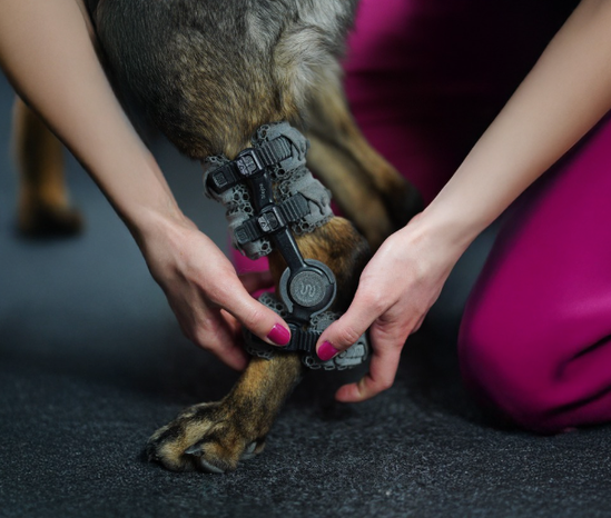
Dog orthotic fittings can become a structured clinical service in a veterinary or rehabilitation practice without requiring the clinic to manufacture devices. The service extends well beyond taking measurements: it includes diagnosis-led patient selection, an appropriate prescription, accurate data capture, professional fitting, owner education, gradual introduction, and planned reassessment.
This practical guide explains how to introduce dog orthotic fittings responsibly, from choosing between WIMBA Go and Pro to evaluating skin, alignment, gait, and functional progress. It also shows how a canine orthotic provider can organise team roles, communicate realistic expectations, and integrate custom dog orthotics into a wider treatment or rehabilitation plan.
Building a Safe Dog Orthotic Fittings Service
A practice should treat each orthosis as a prescribed medical device rather than a retail accessory. The clinical team remains responsible for confirming the diagnosis, defining which movement requires support or control, selecting the treatment objective, checking contraindications, and deciding how the result will be monitored. Current clinical references describe veterinary orthoses as biomechanical tools that must be matched to the individual patient and used with appropriate follow-up.[1][2]
Before accepting the first case, define:
- Who performs the orthopaedic or neurological assessment and confirms the indication
- Who collects product-specific photographs, measurements, scans, and clinical records
- Who conducts the fitting, documents the baseline, and teaches the owner
- How skin concerns, fit problems, damaged components, and urgent questions are escalated
- When routine and outcome-based rechecks are scheduled
- How professional time for assessment, fitting, education, and follow-up is reflected in the service
Professional scope varies between jurisdictions. Clinics should confirm which team members may assess, prescribe, fit, and modify medical devices under local rules. Registration with a manufacturer does not replace a professional licence, clinical competence, or the treating clinician’s duty of care.
Why Offer Custom Dog Orthotics in Your Practice?
Several forces are increasing demand for dog braces for veterinary clinics. The pet population is ageing, and longer lives can bring more degenerative joint disease, neurological presentations, and post-surgical patients requiring extended conservative management. Referral patterns for veterinary rehabilitation services were examined in a cross-sectional survey of 461 US veterinarians, which documented referring clinicians’ perceptions and factors that encouraged or impeded access to rehabilitation facilities.
Post-operative care is another factor. After selected procedures, patients may benefit from external support during a clinician-defined recovery period. A correctly selected and fitted orthosis can help control unwanted movement, support alignment, and facilitate supervised weight-bearing when unrestricted activity would be inappropriate. Device choice and wearing protocol should always reflect the diagnosis, surgery performed, tissue-healing stage, and rehabilitation goal.
Beyond surgery, rehabilitation teams are integrating orthotics into conservative management plans for carpal, tarsal, stifle, and neurological conditions. Owners often research mobility options before arriving at the clinic. A practice that can assess the patient, discuss realistic goals, and coordinate the device pathway is better placed to retain the case and the wider client relationship.
Demand for custom dog orthotics is supported by:
- An ageing pet population with more chronic mobility needs
- Greater access to surgery, rehabilitation, and long-term conservative care
- Growing owner awareness of patient-specific mobility solutions
- Referral demand for clinics able to manage complex fittings and follow-up
- Clinical interest in controlled support during recovery and daily activity
Clinical Outcomes: What Practices Can Evaluate
The clinical rationale for canine orthotics can be organised around three goals: managing mechanical stress, supporting functional mobility, and improving the patient’s ability to participate in an appropriate rehabilitation plan. Veterinary reviews describe orthoses as devices that can restrict, control, or assist motion and protect an affected segment, but outcomes should be defined for the individual case rather than assumed from the device alone.
Mechanical support. A correctly designed orthosis can offload or control structures exposed to excessive stress. In carpal instability, for example, a retrospective series of 14 dogs reported a return to normal function in 79% of patients treated with a custom carpal brace after conservative rest had failed. The treating professional should still determine whether the patient requires sagittal control, multiplanar support, immobilisation, or another intervention.
Functional mobility. The goal of orthotic management is not always complete immobilisation. In a retrospective study of ten dogs with cranial cruciate ligament insufficiency, affected-limb total pressure index improved by 5.1 percentage points after at least 90 days of stifle orthosis use. A computer model also predicted reduced abnormal tibial translation and rotation with a stifle orthosis, while an experimental kinematic study showed that an orthosis altered movement throughout the pelvic limb. These findings support whole-limb gait assessment rather than evaluation of the supported joint in isolation.
Owner-reported quality of life. Owners can provide valuable observations about comfort, confidence, walking tolerance, and participation in daily routines. A comparative study assessed owner satisfaction after management of cranial cruciate ligament disease with a stifle orthosis or TPLO, while a later survey of 56 orthosis users found that owner-reported pet quality of life was associated with the owner’s own reported quality of life and willingness to recommend the device. These subjective reports should complement, rather than replace, clinical reassessment.
The evidence remains limited and heterogeneous. The largest prospective study followed 43 canine orthosis and prosthesis patients, but it had no control group and included different diagnoses and device types. A 2025 report found improved weight distribution in two dogs using custom stifle orthoses, but its very small sample prevents broad conclusions. Clinics offering custom dog orthotics should therefore present the published findings as supportive evidence, not a guarantee of outcome.
Together, these studies justify cautious, measurable treatment goals, transparent owner communication, and scheduled reassessment rather than broad claims about guaranteed clinical success.
A clinic can monitor orthotic outcomes through:
- Joint position and control during standing and walking
- Weight-bearing, gait quality, and activity tolerance
- Skin condition and pressure at the device interface
- Owner adherence and confidence with device application
- Progress toward the documented rehabilitation goal
How Offering Orthotics Differentiates Your Practice
The competitive landscape for general veterinary practice is crowded. Independent clinics may find it difficult to compete on price or convenience alone. Veterinary rehabilitation services built around specialist capability, coordinated follow-up, and genuine clinical depth offer a different form of value that is harder to replicate.
Becoming a canine orthotic provider sends a clear signal to the referral community: the practice has the clinical workflow and supplier support required to manage selected musculoskeletal and neurological cases from assessment through fitting and monitoring. Survey research confirms that referring veterinarians evaluate both the benefits of rehabilitation and practical barriers to accessing a facility, making a clear local pathway relevant to referral development.
There is also a business dimension. Dog braces for veterinary clinics create a professional service pathway that includes assessment, data capture, fitting, client education, and follow-up. The value lies not only in the physical device but also in the clinical expertise and continuity of care around it. Clinics should set fees transparently and explain which appointments, adjustments, and monitoring services are included.
Within veterinary rehabilitation services, orthotics can extend the work completed during an appointment into selected daily activities. The device does not replace therapy, but it may help maintain the prescribed joint position or movement boundary between sessions. This creates a more integrated service and a clearer role for planned reassessment.
Why Clinical Governance Matters
Published evidence supports possible benefits in selected cases but does not justify presenting an orthosis as a universally low-risk solution. A retrospective series of 14 athletic dogs treated for carpal ligament instability reported reduced lameness and return to normal function in 11 dogs; however, there was no untreated control group.
Stifle research includes retrospective gait analysis, biomechanical modelling, kinematic evaluation, owner-reported comparison with TPLO, and a recent two-dog clinical report. The findings are encouraging for selected patients, but they do not demonstrate that bracing is equivalent to surgical stabilisation or appropriate for every dog with cranial cruciate ligament disease.
Complications must be built into the workflow. In a prospective 12-month study, 39 of 43 dogs using an orthosis or prosthesis experienced at least one complication. Skin problems, mechanical issues, and device non-acceptance were the main categories. During the first three months, at least one skin complication occurred in 58% of stifle, 90% of carpal, and 60% of tarsal orthosis cases.
These data do not mean that custom veterinary orthotics should be avoided. They mean the practice needs informed consent, gradual introduction, owner training, rapid access to advice, and scheduled review. A service model that charges only for the device but leaves no time for these tasks is clinically and commercially fragile.
The WIMBA Provider Programme: How It Works
The WIMBA veterinary orthosis programme is designed for veterinary practices, rehabilitation centres, and physiotherapy clinics that want to offer patient-specific devices without building an in-house manufacturing capability. WIMBA handles digital design and production, while the veterinary professional remains responsible for diagnosis, case selection, fitting, client instruction, and clinical follow-up.
The veterinary orthosis programme begins with professional registration and case assessment. Eligible WIMBA Go devices can be ordered in WimbaAPP using the required photographs and manual measurements. WIMBA Pro devices use WimbaSCAN data and require the relevant Provider Pro pathway and scanning tools. This distinction allows the clinical team to select a workflow suited to the condition and case complexity.
WIMBA reviews the submitted patient information and manufactures the selected device to match the supplied anatomy and functional requirements. The fitting takes place through the veterinary professional, who checks alignment, stability, contact areas, gait, and patient response. Ongoing support is available for questions about fit, device configuration, and unusual cases.
Clinics can review the full range of WIMBA devices, use the free case consultation for a current patient, or create an account in WimbaAPP. Case eligibility and the required data should be confirmed before an order is placed. Clinics moving from a supplier that has closed can read what veterinary clinics can do after OrthoPets closed.
- Assess the patient: confirm the diagnosis, affected joint, direction of instability, skin condition, gait changes, and clinical goal.
- Submit the case: share the relevant history, clinical findings, photographs, and imaging where appropriate.
- Select the pathway: determine whether the current Go or Pro criteria and data requirements match the case.
- Place the order: upload the required measurements, photographs, or WimbaSCAN data through WimbaAPP.
- Fit the device: assess alignment and movement, teach application and removal, and introduce the prescribed wearing schedule.
- Monitor the outcome: review skin, fit, gait, activity tolerance, device condition, and progress toward the original goal.
Step 1: Identify Suitable Patients
The first stage of veterinary orthosis fitting is not choosing a product. It is identifying a diagnosed problem that an external device can address. Examination should determine the affected structure, joint stability, planes of abnormal motion, pain, skin condition, neurological status, activity requirements, and concurrent disease. Relevant imaging and surgical history should be reviewed when they affect treatment selection or device design.
Potential candidates may include selected dogs with carpal or tarsal instability, hyperextension, post-operative support needs, or a diagnosed stifle condition for which the clinical team has chosen conservative management or guided rehabilitation. A retrospective series of 14 athletic dogs with carpal ligament instability reported reduced lameness and return to normal function in 11 cases, but the lack of a control group limits the conclusions that can be applied to another patient.[3]
Stifle evidence is also mixed and indication-specific. Published work includes retrospective gait data, biomechanical modelling, kinematic testing in healthy dogs, owner-reported comparisons with TPLO, and a recent two-dog clinical report.[4][5][6][7][10] These studies support careful consideration of bracing in selected cases, not a general claim that an orthosis reproduces surgical stabilisation or eliminates the need for surgery.
Active infection, an unprotected wound, marked dermatitis at the interface, uncontrolled pain, rapidly changing swelling, or anatomy that cannot support the intended device may postpone or prevent fitting. Behaviour, handling tolerance, owner capacity, and access to follow-up also affect candidacy. Tripod status, age, obesity, paresis, or a diagnosis name alone should not automatically trigger an order for custom dog orthotics.
What the Evidence Means for Dog Orthotic Fittings
The available literature supports a cautious, monitored pathway. In a prospective study of 43 dogs using carpal, tarsal, or stifle orthoses or a prosthesis, 39 experienced at least one complication during the 12-month study. Skin problems, mechanical issues, and device non-acceptance were the main categories. During the first three months, at least one skin complication was reported in 58% of stifle cases, 90% of carpal cases, and 60% of tarsal cases.[9]
The same study found objective improvement in many assessed carpal and stifle patients, but there was no control group, so the contribution of the device cannot be isolated from rehabilitation, natural recovery, medication, or other care.[9] This balance is central to responsible dog orthotic fittings: potential functional benefit should be discussed together with adaptation, skin risk, possible repairs, and the need for rechecks.
Owner experience also matters. In a survey of 56 owners, perceived patient quality of life was associated with the owner’s own reported quality of life and willingness to recommend orthotic treatment.[8] Clear education and achievable daily management are therefore clinical parts of veterinary orthosis fitting, not merely customer-service additions.
Practices should collect a baseline and define a measurable goal. Depending on the diagnosis, this may include joint position, controlled range of motion, weight acceptance, gait quality, skin condition, activity tolerance, or an owner-reported pain and function measure. A systematic review found the Canine Brief Pain Inventory, Canine Orthopedic Index, and Liverpool Osteoarthritis in Dogs instrument had sufficient content validity for dogs with osteoarthritis or other joint disease, although the selected measure must suit the patient and intended outcome.[11]
Step 2: Choose the Correct WIMBA Pathway
WIMBA Go and WIMBA Pro use different data-capture workflows. Eligible Go devices are ordered through WimbaAPP using the manual measurements and clear photographs requested for the specific product. They do not require WimbaSCAN. Current Go options and indications should be checked on the WIMBA devices page before the clinic promises a solution.
Pro devices are intended for cases requiring the Pro pathway and use WimbaSCAN together with any required manual measurements and clinical materials. Access requires the applicable WIMBA Provider Pro onboarding and scanning tools. The trained veterinary team captures the scan in the clinical environment; a pet owner should not independently generate or submit the order.
When the indication or pathway is uncertain, use the WIMBA Case Consult before ordering. A useful submission includes the diagnosis, onset and progression, relevant imaging, previous surgery or rehabilitation, patient behaviour, skin concerns, videos or photographs, and a specific functional objective. The resulting device report supports decision-making but does not replace the treating professional’s judgement.
Step 3: Capture Case Data for Custom Dog Orthotics
WimbaAPP is the ordering and case-management platform for veterinary professionals. It guides the user through the materials required for the chosen device and can be used to monitor order progress. Do not reuse a generic measurement checklist across all products: landmarks, limb position, photographs, scan coverage, and supplementary measurements can differ.
Prepare the following before submission:
- Patient signalment, current body weight, body condition, activity, and behaviour
- Confirmed diagnosis, affected side, severity, relevant imaging, and treatment history
- Current skin status, wounds, oedema, pain, neurological findings, and concurrent disease
- A precise functional goal and the movement that should be supported, limited, or guided
- Product-specific measurements, photographs, video, scan, and forms requested in the current workflow
- Contact details for the team member responsible for clarification, fitting, and follow-up
Collect data on a stable, non-slip surface with adequate light and safe patient handling. If pain, anxiety, or movement prevents accurate capture, the clinician should decide whether to pause, change positioning, provide analgesia, or use another appropriate handling plan. Sedation is not an automatic part of dog orthotic fittings; it is a patient-specific medical decision.
Before submitting, check that left and right are unambiguous, measurements use the required units, photographs show the requested landmarks, and the clinical objective is consistent with the selected device. Accurate source data reduce avoidable clarification, but no capture method removes the need for an in-clinic fitting after delivery.
Step 4: Prepare a Dedicated Veterinary Orthosis Fitting
Allow enough uninterrupted time for the first veterinary orthosis fitting, controlled walking, documentation, and owner teaching. A universal 30- or 45-minute appointment cannot be prescribed because complexity, patient behaviour, device type, owner experience, and the number of required adjustments vary. The booking should prevent the fitting from being compressed into an already full routine consultation.
Before application, confirm the patient, device, affected side, orientation, component configuration, intended range of motion, and current prescription. Compare the limb with the condition recorded at data capture. New swelling, a wound, pain, marked weight change, or altered anatomy may require reassessment before the device is used.
Record baseline skin photographs when appropriate, gait or stance video, relevant circumference or weight, and the agreed outcome measure. These materials make later review more objective and help the canine orthotic provider distinguish a fit change from disease progression, owner application error, or a new clinical problem.
Step 5: Apply and Assess the Device
Follow the manual for the exact product and configuration. Do not rely on a universal two-finger strap rule. Correct tension depends on the strap, interface, suspension system, limb shape, and device design. During dog orthotic fittings, the clinical aim is secure positioning without focal hard contact, excessive compression, rotation, distal migration, or loss of the intended joint relationship.
- Apply the device: position liners, sleeves, shells, straps, hinges, and replaceable components exactly as instructed.
- Check standing alignment: confirm that hard elements do not create focal pressure and that the device remains in the intended position.
- Observe controlled movement: evaluate walking, turning, sitting, rising, and any task relevant to the prescription.
- Watch the whole limb: assess paw placement, adjacent joints, compensatory movement, slipping, noise, and patient behaviour.
- Remove and inspect: compare the skin with baseline and document any pressure mark, heat, swelling, moisture, abrasion, or pain.
- Retest after correction: repeat the relevant checks whenever application or configuration changes.
A dog bearing weight immediately does not prove that the fit is correct, and initial hesitation does not automatically mean the device has failed. Interpret the response together with alignment, skin, gait, diagnosis, pain, and behaviour. Persistent distress, worsening lameness, toe dragging, collapse, rotation, or refusal to move requires reassessment before the patient leaves.
Step 6: Teach Safe Introduction and Daily Management
The owner should demonstrate application and removal while the clinical team observes. Correct errors before discharge and provide the current product manual. A clinic may also record a patient-specific application video with the owner’s consent, but the recording should supplement rather than replace written stop criteria and professional contact information.
Do not prescribe a generic starting period of 30–60 minutes or assume every patient reaches full activity within two weeks. Current WIMBA limb-device manuals commonly begin with short controlled walks, for example up to 15 minutes no more than three times daily in the first week, followed by gradual progression when skin, fit, gait, and behaviour remain acceptable. The exact device manual and clinician-directed schedule take priority.
Owners should remove the device as instructed, inspect all contact areas, keep the orthosis clean and dry, and supervise the patient throughout use. Current WIMBA fitting guidance advises removal if swelling, irritation, pressure sores, or wounds appear; it also warns against continuous 24-hour wear, leaving the patient unattended, using harsh chemicals, machine drying, or altering the structure.
Give the owner a clear escalation route. Persistent redness, heat, swelling, hair loss, abrasion, discharge, pain, licking, device rotation, slipping, damaged parts, unusual sound, new reluctance, or worsening gait should prompt removal and contact with the clinic. Owner confidence improves when a canine orthotic provider explains both the purpose of each component and the signs that require action.
Step 7: Schedule Rechecks and Measure Outcomes
There is no universal evidence-based rule that every patient must return at seven or ten days. Arrange an early review according to the device, skin risk, diagnosis, owner confidence, patient behaviour, and wearing schedule, then plan further reviews around clinical progress. Give owners an earlier route when a concern appears.
At each review, inspect the skin and device, repeat relevant fit and gait checks, confirm application technique, and compare the result with the original goal. Record body weight, muscle or limb-volume change, activity, component wear, adherence, and any periods without use. Do not rely on an unsupported 10–15% circumference threshold; even a smaller local change may matter at a sensitive interface.
Custom dog orthotics may need configuration changes, replaceable components, authorised adjustment, repeat data capture, or a new prescription as the patient changes. The WIMBA Provider should document the concern with the media requested by support and avoid grinding, heating, drilling, cutting, or otherwise altering the structural shell unless explicit case-specific instructions have been provided.
Outcome review should answer a clinical question: is the intended movement better controlled, is the patient using the limb more appropriately, has activity tolerance changed, and are adverse effects acceptable? If the original goal is not being met, reconsider diagnosis, fit, wearing schedule, rehabilitation, pain management, owner application, or whether the device remains appropriate.
Common Challenges with Custom Dog Orthotics
Skin irritation or a pressure area: remove the device, document the location and appearance, assess whether the cause is fit, application, contamination, moisture, activity, or a clinical change, and contact support when needed. Do not add padding automatically because it can transfer pressure or make the interface tighter.
Rotation, slipping, or loss of alignment: verify orientation, strap sequence, tension, liners, component position, and current limb shape. Persistent movement may require an authorised adjustment or design review rather than progressively tighter straps.
Difficulty applying the device: repeat hands-on teaching, simplify the written sequence, label components if approved, and review a patient-specific video. The owner should be able to reproduce correct application before unsupervised home management begins.
Patient non-acceptance: first exclude pain, poor fit, neurological deterioration, fear, and an inappropriate task. A clinician-directed behaviour plan may use brief, positive exposure, but no fixed adaptation time or guaranteed success should be promised. Prospective data show that non-acceptance is a genuine possible complication.[9]
Anatomical change: growth, oedema reduction, weight change, muscle gain or loss, disease progression, and surgery can alter the interface. Reassess rather than assuming that a device remains suitable because its shell is undamaged.
Calculate the Real Cost of the Service
The business case for custom dog orthotics should be calculated from the clinic’s own costs, capacity, and market. There is no evidence that simply adding orthotics guarantees revenue, retention, referrals, or improved outcomes. A sustainable model prices the complete professional pathway and leaves capacity for follow-up and troubleshooting.
Include these elements in the financial model:
- Clinical assessment, record review, imaging interpretation, and treatment planning
- Case consultation, photography, measurements, scanning, forms, and submission time
- Device purchase, shipping, taxes, payment fees, and exchange-rate exposure where relevant
- Dedicated fitting, owner education, written materials, and baseline documentation
- Included and separately billable rechecks, troubleshooting, and communication time
- Pro onboarding, equipment, training, maintenance, and staff capacity when applicable
- Allowance for cancelled appointments, remakes, non-acceptance, and unrecoverable professional time
Decide what the quoted package includes. Owners should know whether assessment, the device, fitting, components, one or more reviews, and rehabilitation sessions are included or charged separately. Pricing must comply with local tax, professional, consumer, and advertising rules.
A WIMBA Provider should also define its policy for deposits, order approval, cancellations, anatomical change after data capture, and manufacturer-supported adjustments. Clear terms reduce conflict without shifting inappropriate clinical or legal risk to the owner.
Review the cost model for custom veterinary orthotics whenever supplier terms, staffing, or follow-up demand changes.
Measure Whether the Service Is Viable
Start with a defined review period rather than assuming demand. Track enquiries, suitable cases, consultations, orders, cancellations, fitting time, unplanned contacts, rechecks, complications, contribution margin, and clinical outcomes. Separate Go and Pro because their acquisition, equipment, and staff-time profiles differ.
Monitor conversion responsibly. A declined device is not automatically a sales failure; the patient may be unsuitable, surgery may be preferable, or the owner may be unable to manage daily care. The purpose of a veterinary orthotics service is to identify and support appropriate cases, not maximise device volume.
Review outcomes alongside operational data. Owner satisfaction is relevant because the owner performs daily management. In a survey of 56 owners, perceived patient quality of life was associated with the owner’s own reported quality of life and willingness to recommend the experience. This supports clear expectation-setting, but it does not prove that a dog orthotic service will improve clinic retention.
After the review period, decide whether to continue, modify, or expand the service. Expansion may involve team training, protected appointment slots, relationships with referring clinics, rehabilitation capacity, or progression from Go to Pro. Use observed demand and workload rather than optimistic assumptions.
Before expanding the veterinary orthotics service, confirm that the dog orthotic service remains clinically manageable.
A Practical Launch Checklist
- Review local professional scope, consent, advertising, pricing, and recordkeeping requirements.
- Create a verified WimbaAPP account and decide whether the clinic will begin with Go only or enter the Pro pathway.
- Name the clinical lead and assign assessment, data capture, fitting, owner training, and support roles.
- Build case-selection, submission, fitting, adverse-event, and recheck templates.
- Set package inclusions, professional fees, deposits, cancellation terms, and review costs.
- Test the workflow with selected cases and schedule enough time for education and follow-up.
- Measure workload, outcomes, complications, owner experience, and contribution margin before expanding.
For a first or uncertain case, a WIMBA Provider can submit clinical information for review before ordering. Early consultation is particularly useful when anatomy, severity, skin condition, previous surgery, or the functional objective makes the pathway unclear.
Workflow for a Canine Orthotic Provider
A repeatable pathway makes dog orthotic fittings easier to deliver consistently. Use templates for assessment, informed consent, baseline media, fitting checks, owner instructions, adverse-event documentation, and rechecks. Store the current device manual with the patient record and record the exact configuration supplied.
Consistent records also make handovers safer when several team members participate.
Eligible Go cases can be ordered through WimbaAPP by a WIMBA Provider without Pro scanning equipment. Clinics that want access to Pro devices need the current WIMBA Provider Pro pathway and WimbaSCAN tools. This distinction should be built into training, appointment planning, and pricing before the service is promoted.
A veterinary orthosis fitting can be offered within a rehabilitation programme or coordinated with another treating professional. Whether physiotherapy, hydrotherapy, medication, surgery, weight management, or home exercise is included depends on the diagnosis and goal. An orthosis should not be marketed as independently superior to combined care when comparative evidence is unavailable.
Client communication should remain functional and specific: explain what the device is intended to support, what it cannot repair, when it will be worn, how progress will be judged, and what complications are possible. This approach helps a canine orthotic provider build confidence without guaranteeing comfort, avoiding surgery, or returning to a particular activity.
Frequently Asked Questions About Dog Orthotic Fittings
Does a canine orthotic provider need a specialist qualification?
WimbaAPP is available to verified veterinary professionals for eligible Go orders, while Pro devices require the current Provider Pro onboarding and scanning tools. Professional scope differs by jurisdiction, so each clinic must confirm who may assess, prescribe, fit, and manage a medical device under local rules.
What is the difference between WIMBA Go and Pro?
Eligible Go devices use the product-specific manual measurements and photographs requested in WimbaAPP and do not require WimbaSCAN. Pro devices require the applicable WIMBA Provider Pro pathway, scanning tools, a veterinary-team scan, and any additional measurements or clinical materials requested.
How do I order custom dog orthotics?
Assess the patient, confirm the indication and goal, select the correct Go or Pro pathway, and submit the required case data through WimbaAPP. If device selection is uncertain, request a WIMBA Case Consult before ordering.
What should happen during a veterinary orthosis fitting?
The clinic should verify the patient, side, device orientation, alignment, contact areas, suspension, and configuration; observe controlled movement; compare skin before and after use; document the baseline; and teach the owner safe application, removal, monitoring, and care.
How quickly should wearing time increase?
Follow the current manual for the exact device and the clinician’s patient-specific plan. WIMBA limb-device manuals commonly begin with short controlled walks and gradual progression, but skin, fit, gait, behaviour, diagnosis, and treatment goals determine whether progression is appropriate.
What if the orthosis does not fit correctly?
Stop progression, remove the device if skin, pain, or gait is concerning, reassess application and current anatomy, document the issue, and contact WIMBA support. Do not structurally modify the device without explicit case-specific authorisation.
Can orthotic fitting be offered outside rehabilitation?
It can be organised as a defined clinical service, but the patient may still need coordinated surgery, medication, rehabilitation, weight management, or another intervention. The diagnosis and functional goal determine whether the orthosis is one component of wider care.
Medical Disclaimer: This material is intended for veterinary professional education and does not replace patient-specific examination, diagnosis, informed consent, prescribing, or treatment planning. Device selection, data capture, fitting, wearing schedules, rehabilitation, and follow-up should be directed by appropriately qualified professionals working within local regulations. Product indications, availability, processes, and timelines may change; confirm current WIMBA guidance before ordering. No specific clinical outcome is guaranteed.
Scientific References
- Wendland T, Hart J, Mich PM, Moe J. Veterinary orthotics, prosthetics, and assistive devices. In: Zink C, Van Dyke JB, editors. Canine Sports Medicine and Rehabilitation. 2nd ed. Wiley; 2025. doi:10.1002/9781394251452.ch15
- Marcellin-Little DJ, Drum MG, Levine D, McDonald SS. Orthoses and exoprostheses for companion animals. Vet Clin North Am Small Anim Pract. 2015;45(1):167–183. doi:10.1016/j.cvsm.2014.09.009
- Tomlinson JE, Manfredi JM. Evaluation of application of a carpal brace as a treatment for carpal ligament instability in dogs: 14 cases (2008–2011). J Am Vet Med Assoc. 2014;244(4):438–443. doi:10.2460/javma.244.4.438
- Carr BJ, Canapp SO Jr, Meilleur S, Christopher SA, Collins J, Cox C. The use of canine stifle orthotics for cranial cruciate ligament insufficiency. Veterinary Evidence. 2016;1(1). doi:10.18849/ve.v1i1.10
- Bertocci GE, Brown NP, Mich PM. Biomechanics of an orthosis-managed cranial cruciate ligament-deficient canine stifle joint predicted by use of a computer model. Am J Vet Res. 2017;78(1):27–35. doi:10.2460/ajvr.78.1.27
- Torres BT, Fu YC, Sandberg GS, Budsberg SC. Pelvic limb kinematics in the dog with and without a stifle orthosis. Veterinary Surgery. 2017;46(5):642–652. doi:10.1111/vsu.12634
- Hart JL, May KD, Kieves NR, Mich PM, Goh CSS, Palmer RH, Duerr FM. Comparison of owner satisfaction between stifle joint orthoses and tibial plateau leveling osteotomy for the management of cranial cruciate ligament disease in dogs. J Am Vet Med Assoc. 2016;249(4):391–398. doi:10.2460/javma.249.4.391
- Lee S, Wendland TM, Rao S, Magee C. Orthotic device use in canine patients: owner perception of quality of life for owners and patients. Front Vet Sci. 2021;8:709364. doi:10.3389/fvets.2021.709364
- Rosen S, Duerr FM, Elam LH. Prospective evaluation of complications associated with orthosis and prosthesis use in canine patients. Front Vet Sci. 2022;9:892662. doi:10.3389/fvets.2022.892662
- Muršec A, Pavlović M, Lampe T, Erjavec V. The impact of knee orthoses on lameness and weight distribution in canine after rupture of the cranial cruciate ligament. Animals. 2025;15(4):545. doi:10.3390/ani15040545
- Radke H, Joeris A, Chen M. Evidence-based evaluation of owner-reported outcome measures for canine orthopedic care: a COSMIN evaluation of 6 instruments. Veterinary Surgery. 2022;51(2):244–253. doi:10.1111/vsu.13753