 Skip to content

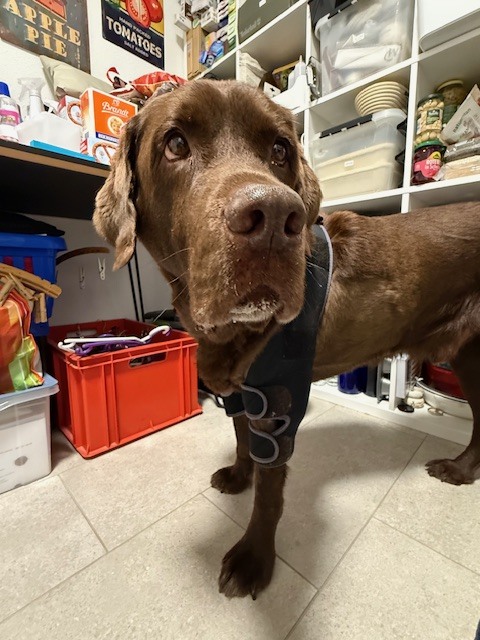
Wound complications in dogs are not always caused by a poor choice of topical dressing. Persistent pressure, friction, movement, moisture imbalance, infection, devitalised tissue, systemic disease, and an unsuitable bandage construct can all prevent orderly healing. When mechanical loading is part of the problem, the wound plan must protect the primary dressing and reduce repeated contact without creating a new pressure point.
This clinical guide explains how to recognise pressure-related risk, where protective wound care spacers may fit, and how to use WIMBA Wound Care without overstating the evidence. The product is an adjunct to professional wound assessment and bandaging, not a replacement for debridement, infection control, surgery, repositioning, or a suitable primary dressing.
Why Wound Complications in Dogs Need Mechanical Assessment
Normal healing progresses through overlapping inflammatory, repair, and maturation phases. A structured wound review can use the TIME framework: tissue, inflammation or infection, moisture, and the wound edge or epithelial advancement. The framework helps clinicians identify why progress has stalled, but it must be interpreted alongside perfusion, pressure, pain, movement, patient health, and the treatment already in use.[1]
Pressure matters because sustained external loading can impair local blood flow, particularly where soft-tissue coverage is limited. Friction and shear can add tissue distortion, while excessive moisture can weaken surrounding skin. These mechanisms may coexist, but they should not be assumed from appearance alone. Infection, foreign material, neoplasia, vascular compromise, metabolic disease, malnutrition, self-trauma, and instability can produce similar delayed-healing patterns.
Before changing the dressing plan, document:
- Wound cause, location, dimensions, depth, tissue type, exudate, odour, pain, and surrounding skin
- Perfusion, oedema, neurological function, mobility, body condition, and relevant systemic disease
- Contact with floors, bedding, casts, splints, orthoses, bandage edges, or another external surface
- Change in size and appearance using repeatable photographs and measurements
- The clinical objective of every dressing layer and any proposed offloading component
Good veterinary wound management therefore begins with a diagnosis and a measurable objective. A spacer is appropriate only when creating clearance or redistributing external forces addresses an identified part of the problem.
What the Veterinary Evidence Says About Pressure
Direct canine evidence confirms that pressure is clinically relevant, although it does not establish one universal safe threshold. In a study using three canine cadavers, recurrent high-risk contact zones in lateral recumbency included the scapulohumeral region, greater trochanter, and thirteenth rib. Pressure-relieving mats reduced measured pressure, but static support alone did not consistently maintain the study’s proposed safe level, especially in thin dogs. The authors therefore recommended frequent repositioning.[2]
Canine decubital ulcers can become chronic. A randomised blinded trial involving 18 dogs with bilateral chronic ulcers over the greater trochanter or shoulder illustrates the severity and persistence of this disease, although the trial evaluated platelet gel rather than a mechanical spacer.[3] It supports treating the biological wound bed while the source of repeated pressure is also addressed.
External coaptation research shows why adding material without monitoring can be dangerous. In a retrospective study of 60 dogs and cats treated with casts, 63% developed a soft-tissue injury; clinicians, rather than owners, identified 80% of those injuries.[4] A separate series of nine dogs and two cats described severe ischaemic bandage injuries, including cases requiring grafting, digit amputation, or limb amputation.[5]
More recent measurements reinforce the need for repeat assessment. Reapplication of the same rigid fibreglass splint by a second clinician produced a change of at least 20 mmHg at one or more measured sites in 10 of 11 dogs, usually as an increase; pressure also changed unpredictably between recumbency and standing.[6] In 13 dogs, a shorter pelvic-limb cast generated greater peak pressure at the calcaneus and proximal cast edge than a taller configuration.[7]
These studies do not test WIMBA Wound Care and should not be presented as product efficacy trials. They demonstrate that contact pressure, anatomy, construct design, position, and reapplication can change tissue loading. That is the clinical rationale for deliberate pressure offloading for dogs, followed by direct skin and wound inspection.
How Mechanical and Biological Factors Interact
Pressure: prolonged loading over a bony prominence or beneath a rigid edge may impair perfusion and cause local tissue damage. The risk depends on magnitude, duration, tissue condition, anatomy, and the patient’s ability to move. Padding is not automatically protective; canine research has shown that placement and geometry influence pressure over prominences.[8]
Friction and shear: rubbing acts at the surface, while shear distorts deeper tissues as adjacent layers move differently. A spacer may reduce direct contact at a selected site, but no spacer can eliminate all movement within a bandage or compensate for poor retention, unsuitable activity, or a slipping construct.
Moisture and exudate: a moist wound environment can support healing, whereas uncontrolled exudate and prolonged wetness can damage periwound skin. An open lattice may support airflow around the protected area, but exudate management remains the job of the wound-contact layer, absorbent materials, change schedule, and clinical treatment.
Inflammation, infection, and systemic factors: bacterial burden, necrotic tissue, foreign material, immunosuppression, endocrine disease, nutrition, perfusion, pain, and medication can alter healing. Mechanical protection cannot diagnose or correct these factors. Each must be managed within the wider canine wound care plan.
Patients That Need Closer Pressure Surveillance
Risk should be assessed individually rather than assigned from diagnosis alone. Wound complications in dogs are more likely to involve repeated contact when the patient cannot reposition normally, the lesion lies over a prominence, or an external device changes local loading.
Consider enhanced surveillance for:
- Recumbent, sedated, frail, or severely mobility-limited patients
- Thin dogs with limited soft-tissue coverage over bony prominences
- Dogs with reduced sensation, paresis, paralysis, or altered protective responses
- Patients wearing casts, splints, orthoses, or long-term bandages
- Postoperative wounds near joints, distal limbs, or areas exposed to repeated contact
- Existing calluses, pressure sores, fragile scars, oedema, or compromised periwound skin
Tripod status may change loading across the remaining limbs, but it does not by itself prove that a spacer is needed. Examine the actual contact point, gait, resting posture, skin, and wound. The broader biomechanical considerations for three-legged dogs are covered in our tripod dog resources.
When Wound Care Spacers May Be Appropriate
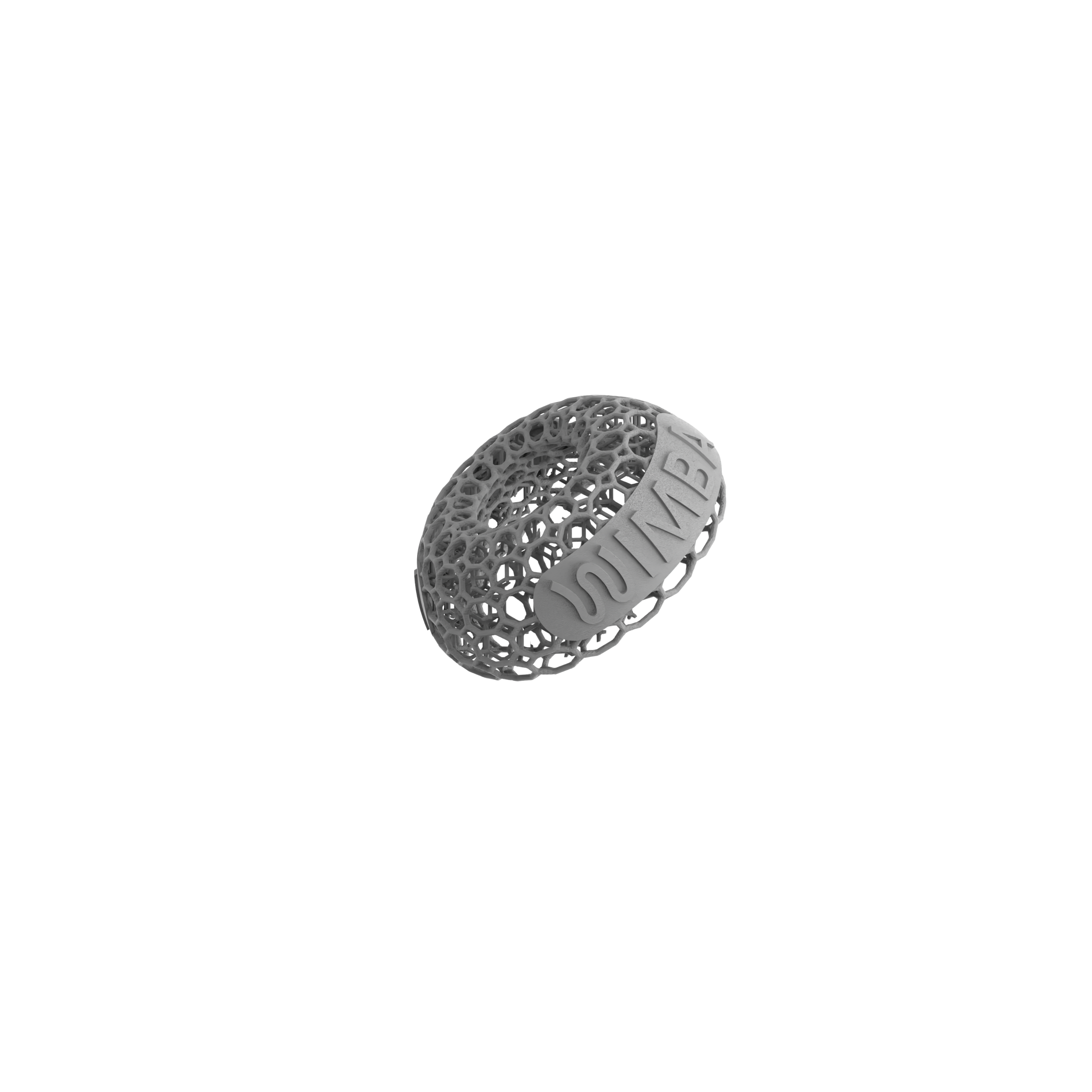
A protective spacer is considered when the team needs to create clearance over a selected site, reduce direct contact, or distribute an external force across a wider area. Potential applications include focal wounds, pressure sores, calluses, sutured areas, and linear incisions that are otherwise exposed to repeated contact. Product choice depends on wound geometry, location, surrounding tissue, patient position, and the complete bandage design.
The spacer must not contact an open wound. Current WIMBA instructions place the selected component over a standard primary wound cover, then secure it within an outer bandage layer. The primary dressing protects and manages the wound surface; the spacer provides external mechanical protection.
Do not use wound care spacers to mask an unknown cause of deterioration. Progressive necrosis, unexpected pain, purulent discharge, malodour, dehiscence, exposed deep structures, systemic illness, or loss of limb perfusion requires prompt reassessment and treatment directed at the underlying problem.
How WIMBA Wound Care Fits the Treatment Plan
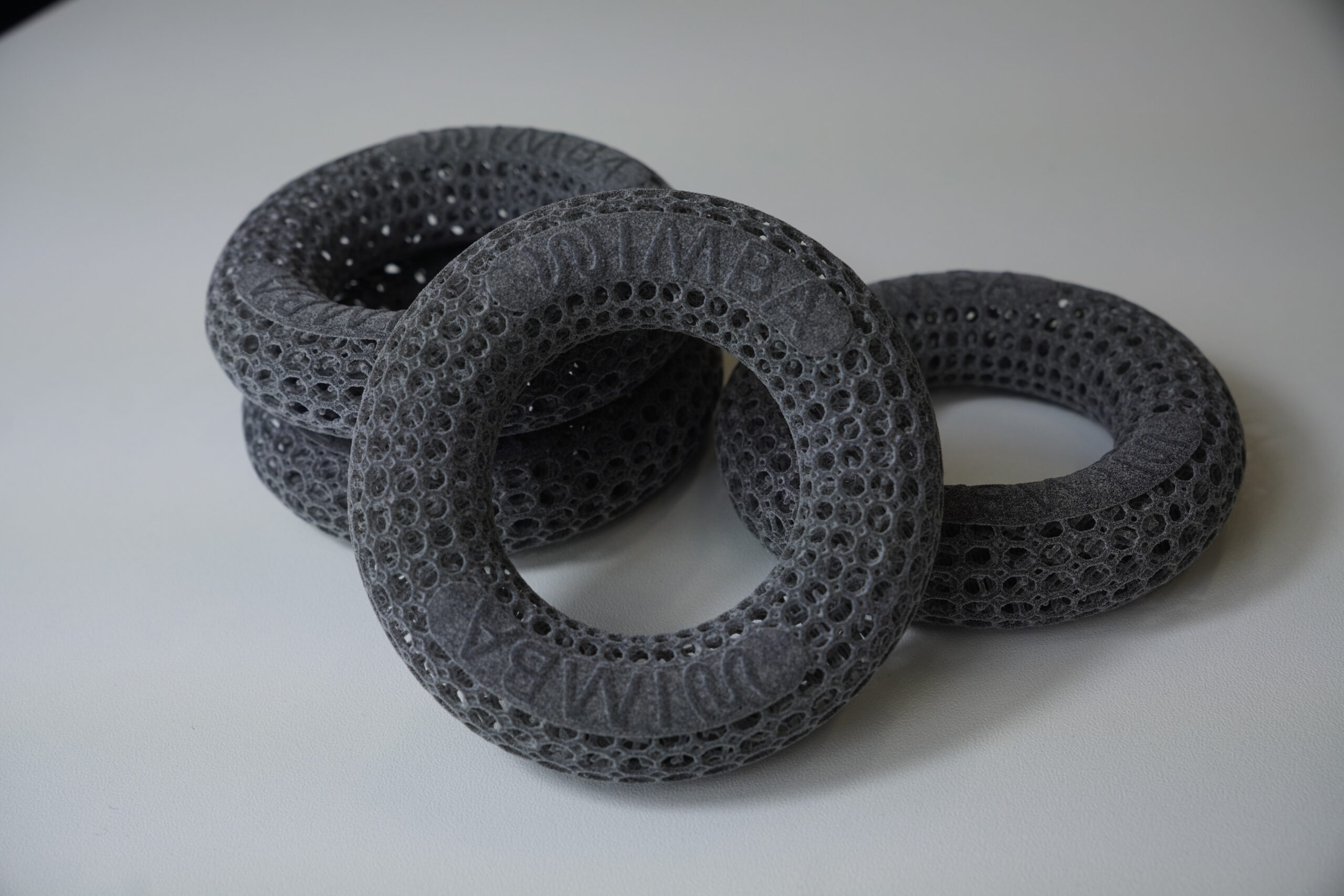
The current range consists of standard 3D-printed protective components rather than patient-specific devices manufactured from a scan. Clinics select among available forms and sizes according to the wound site and the required type of protection. No WimbaSCAN, cast, or custom engineering submission is needed for these products.
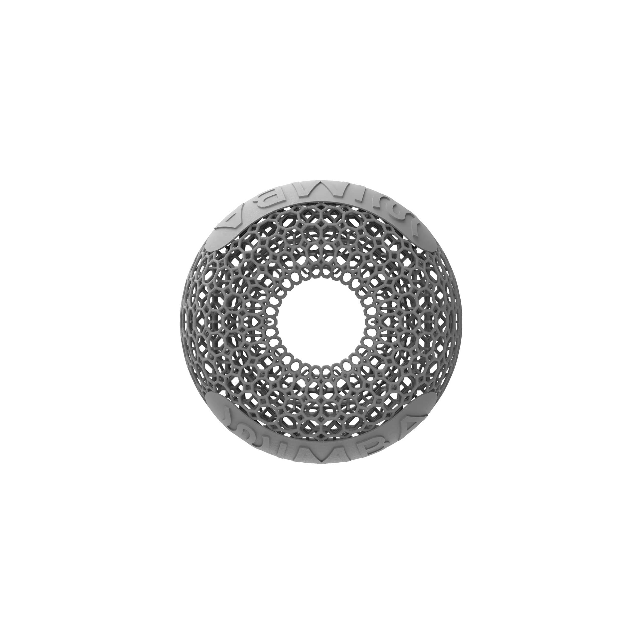
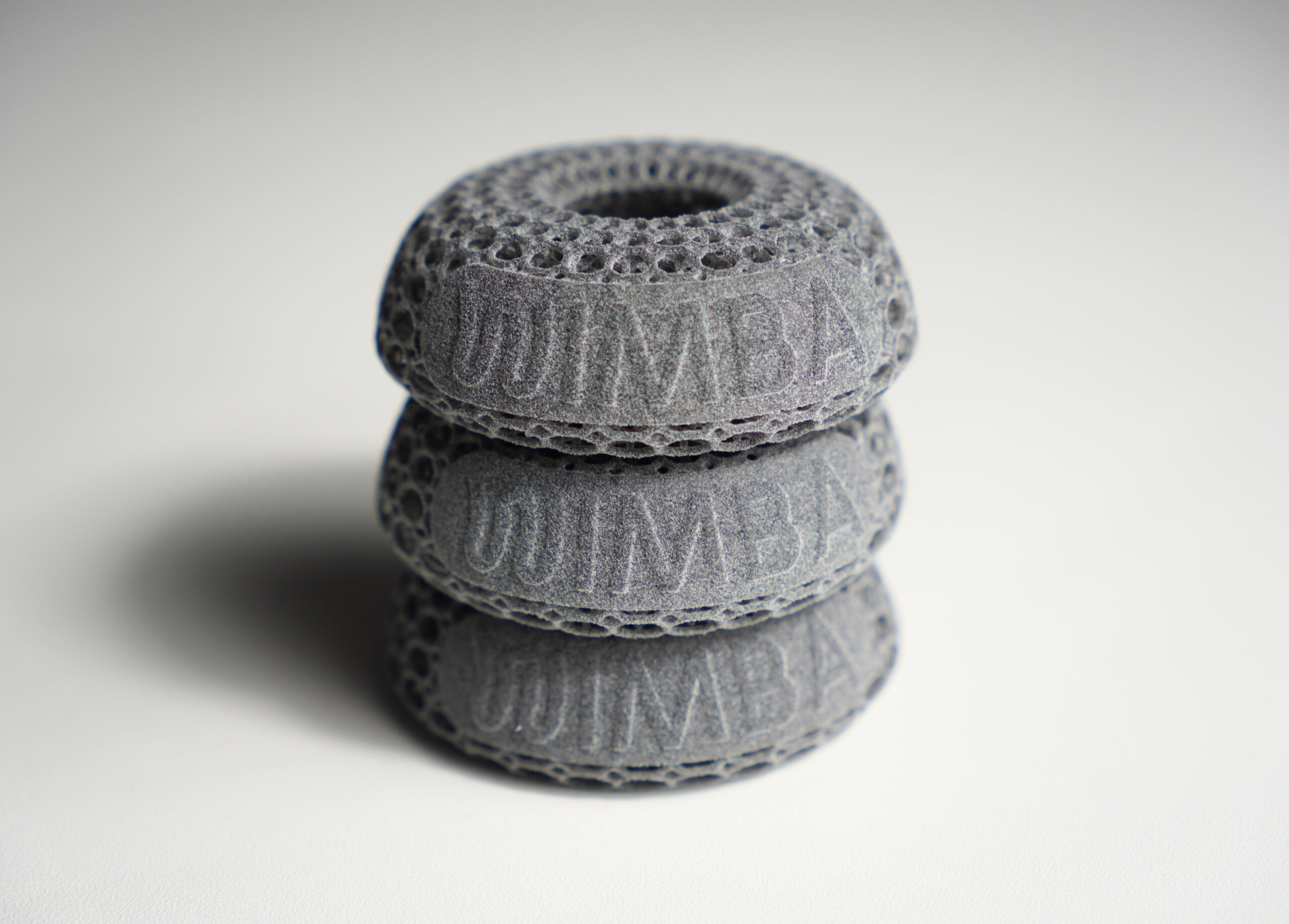
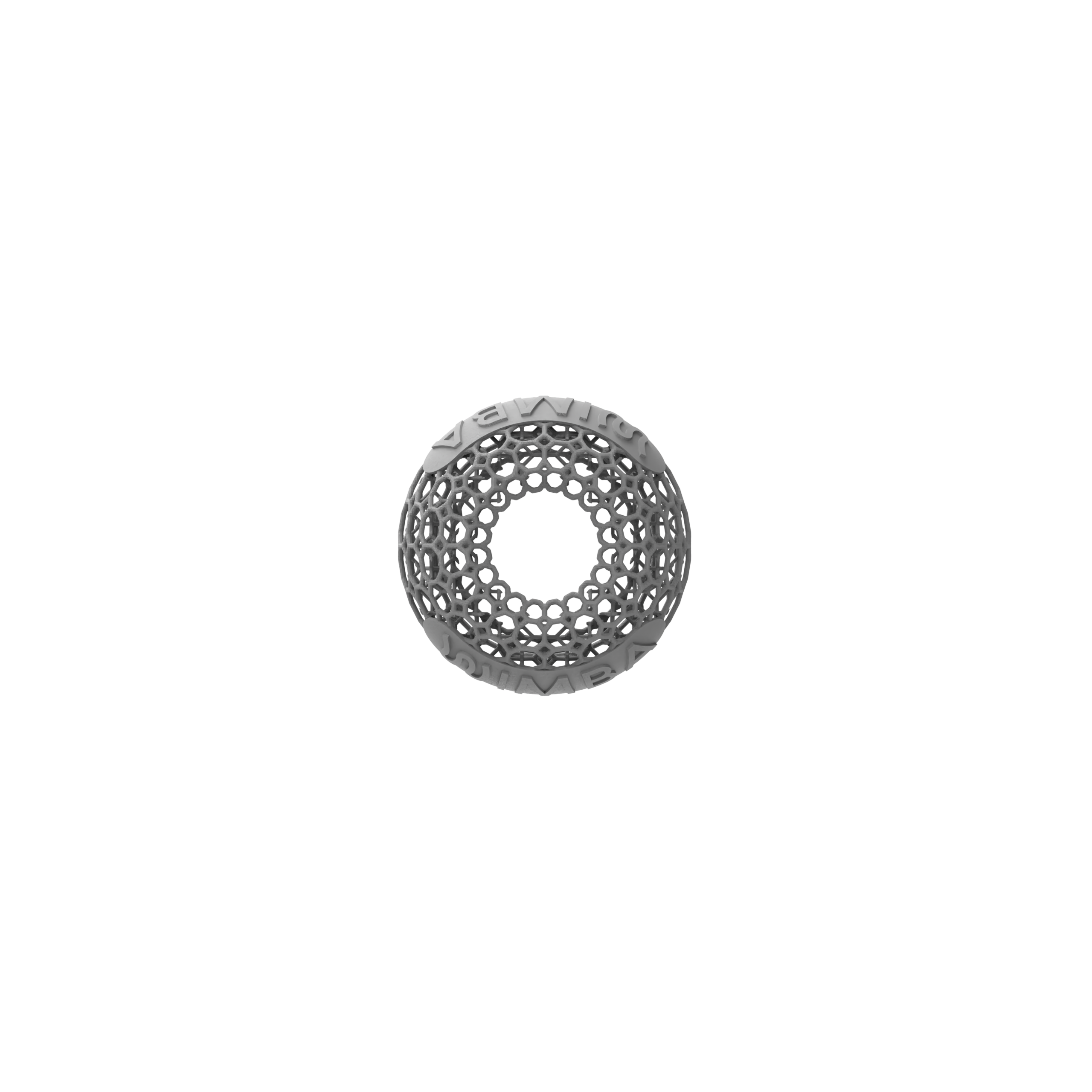
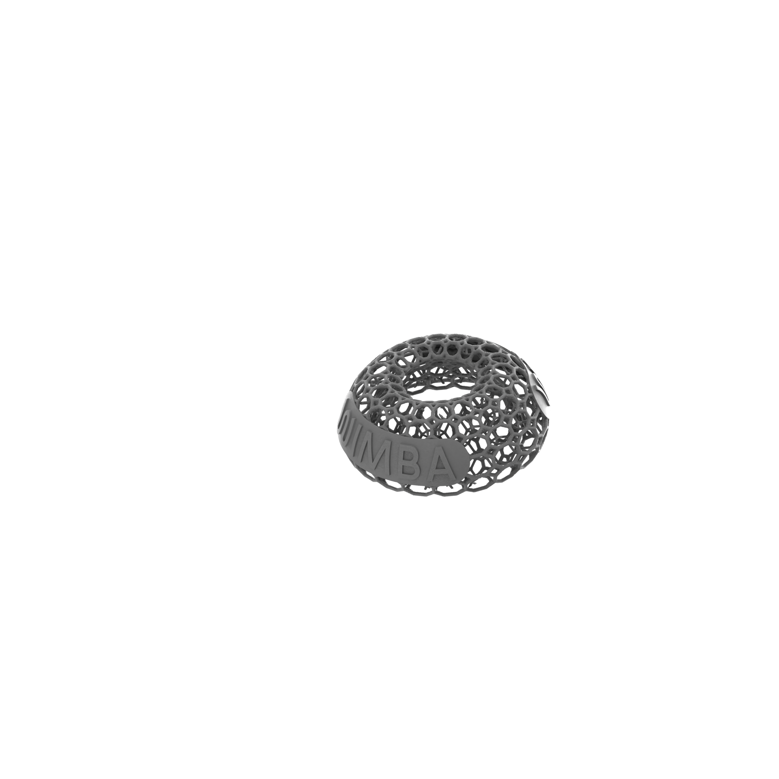
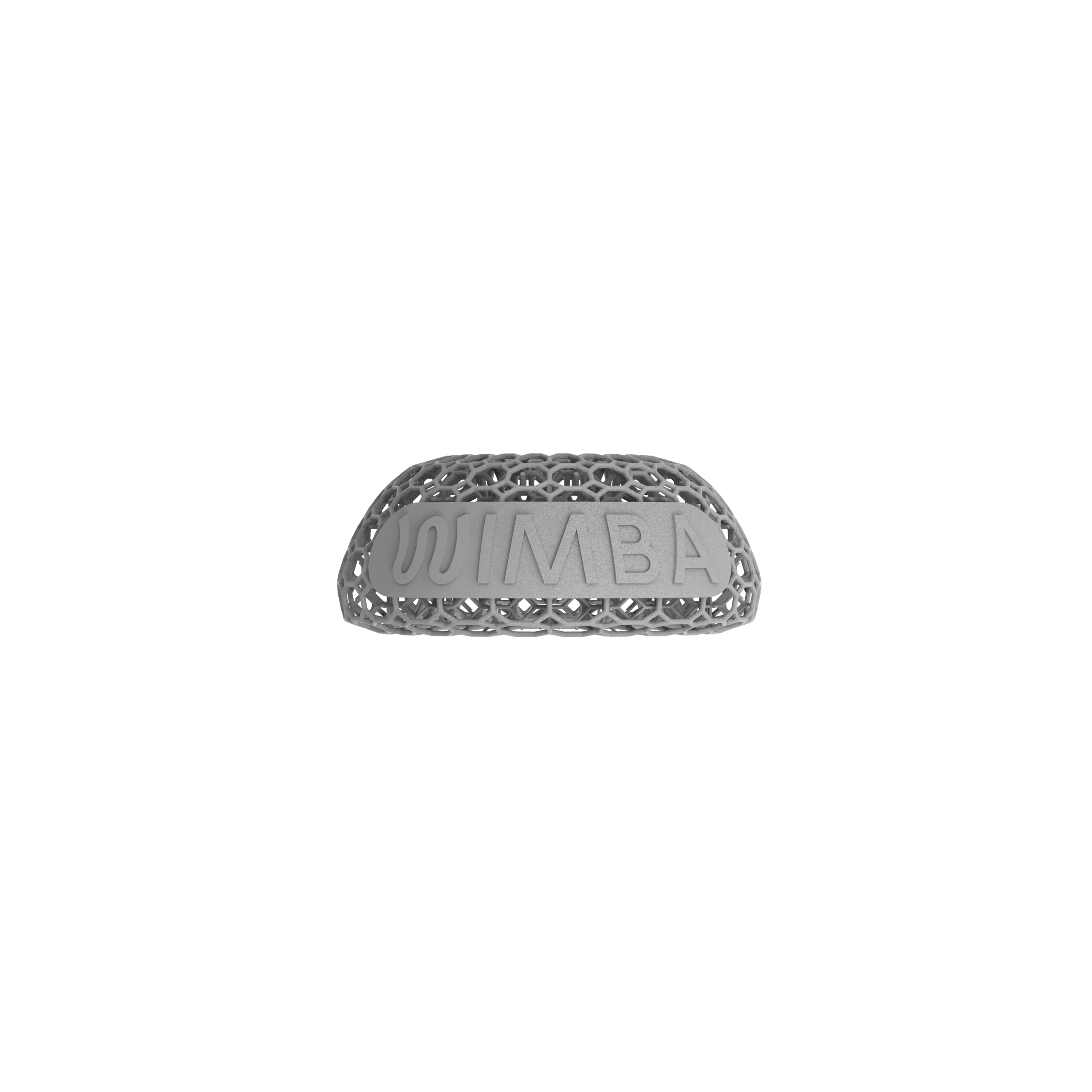
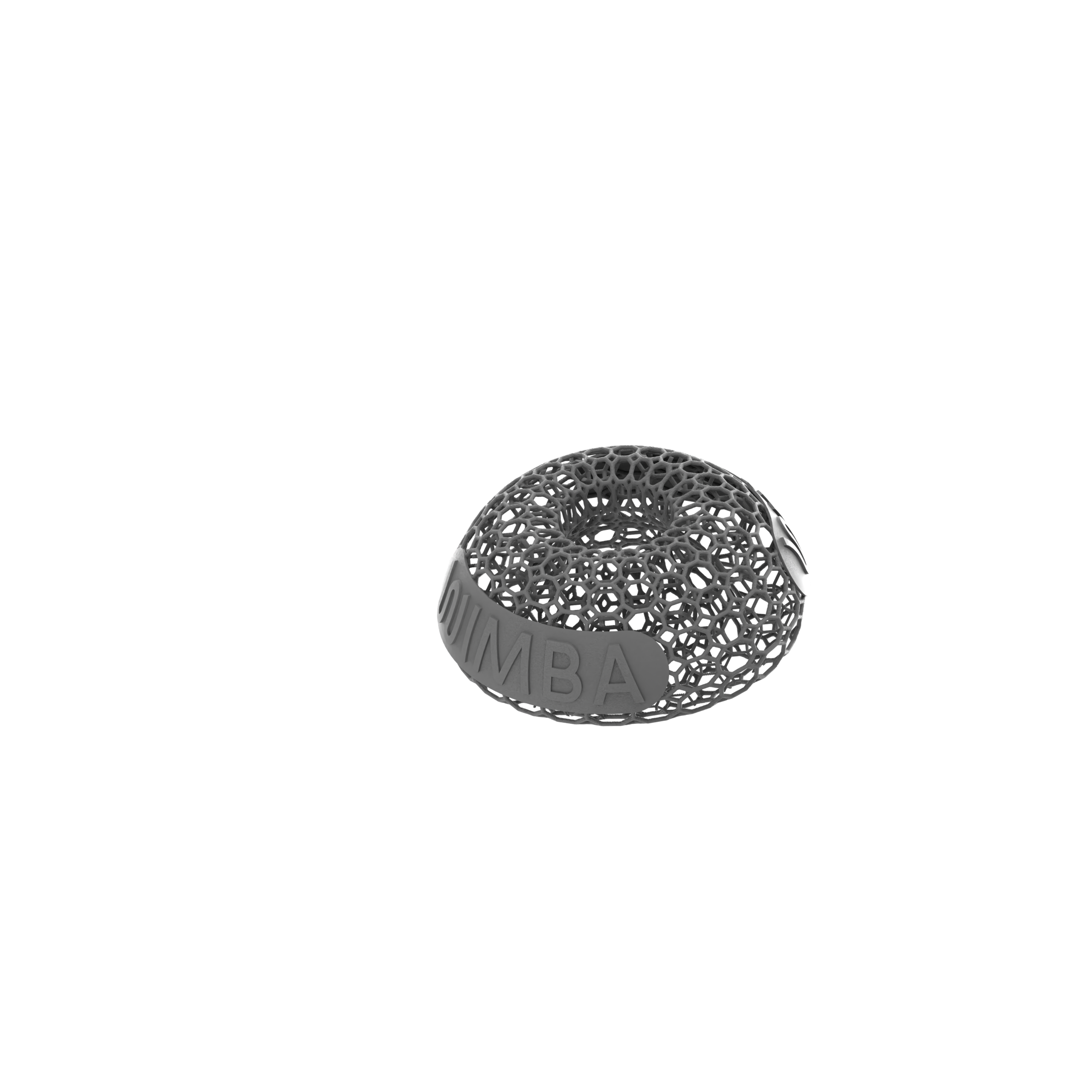
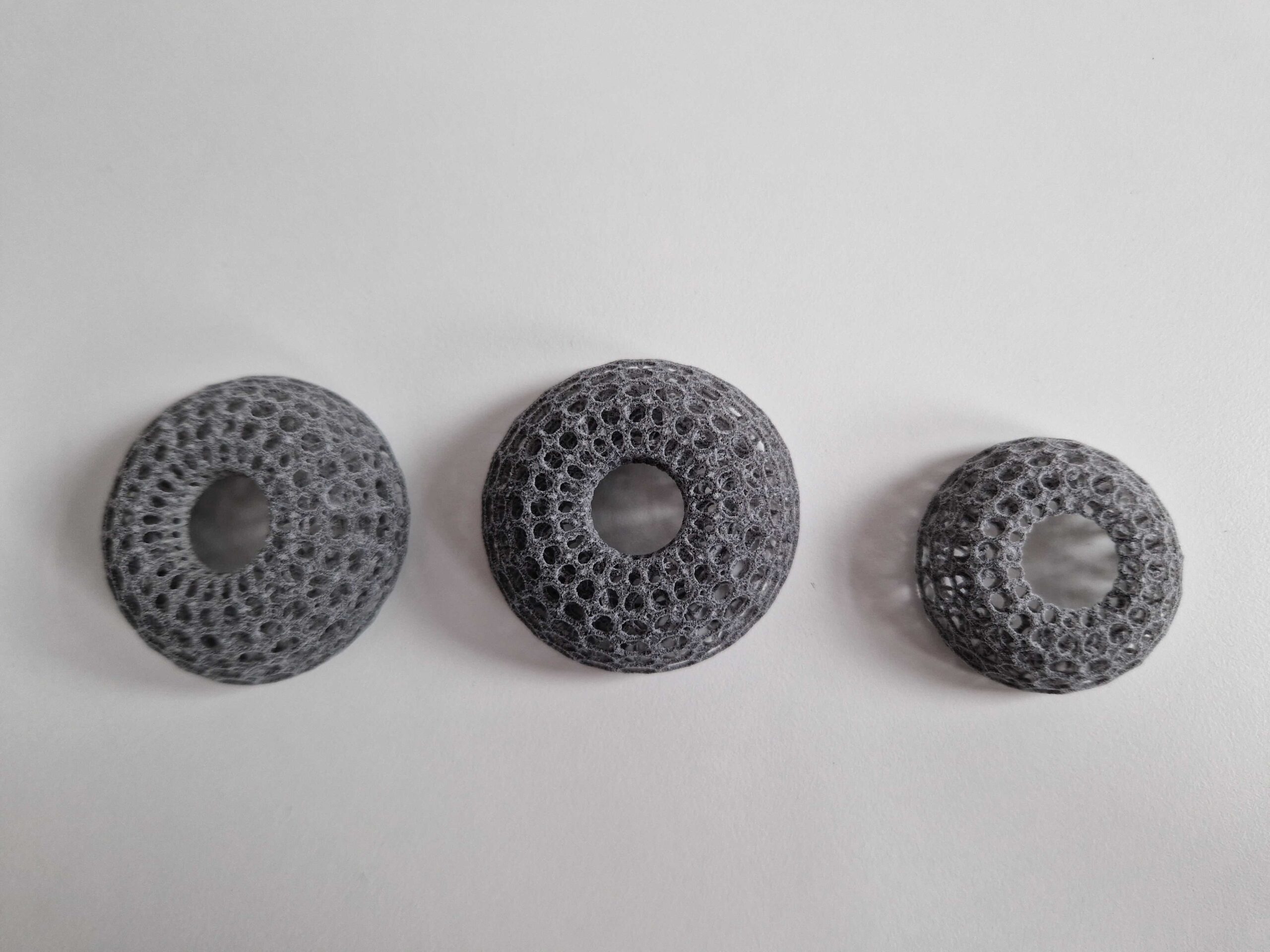
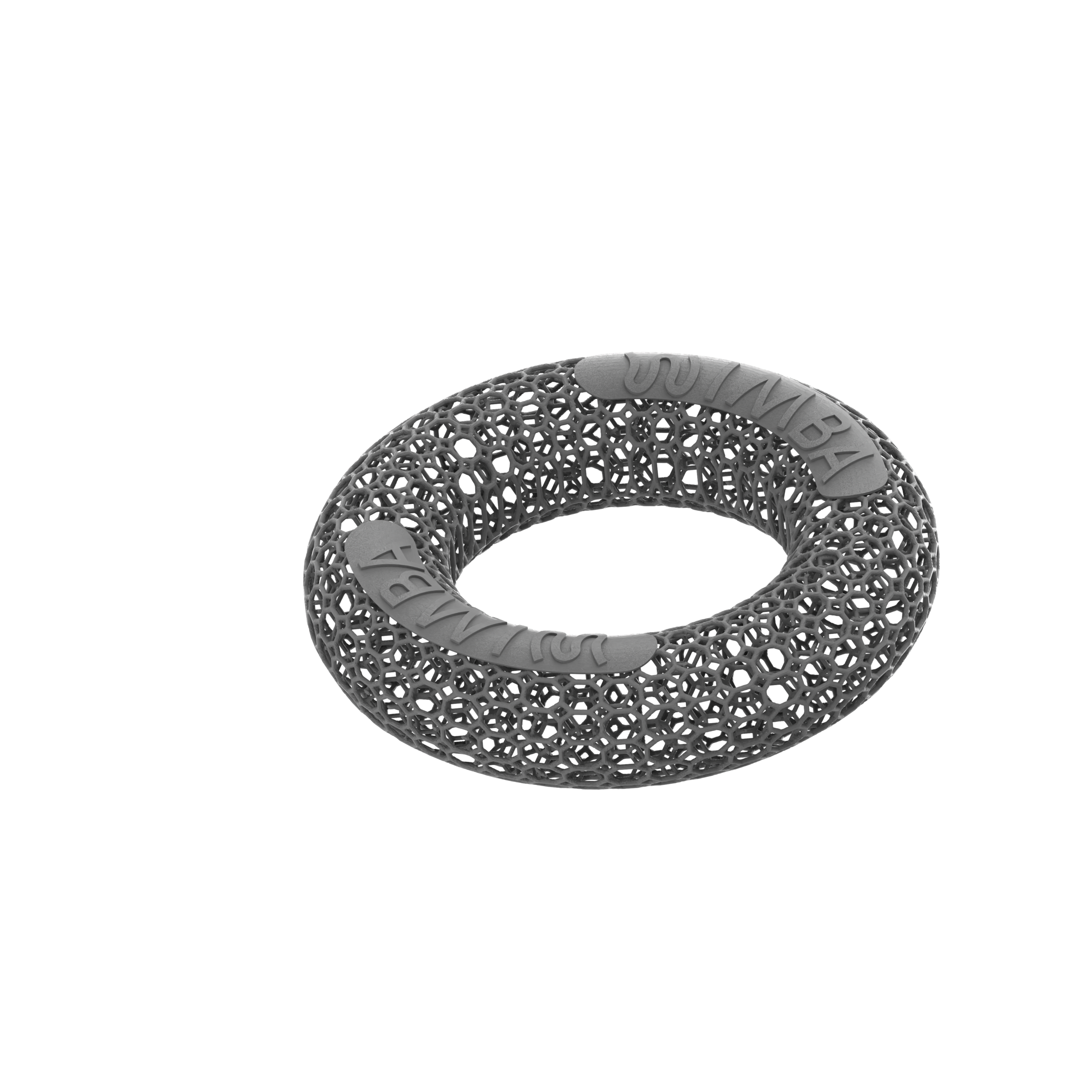
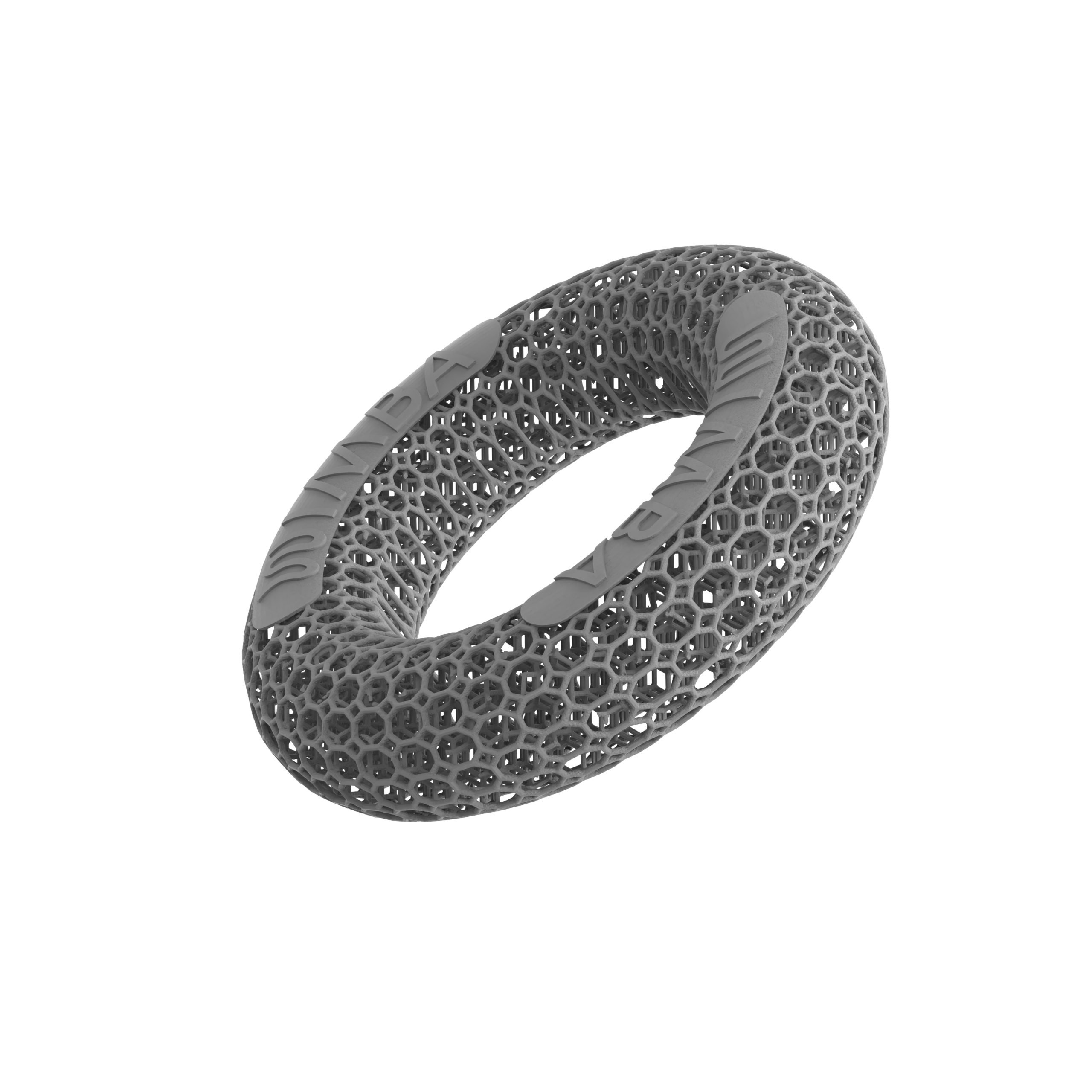
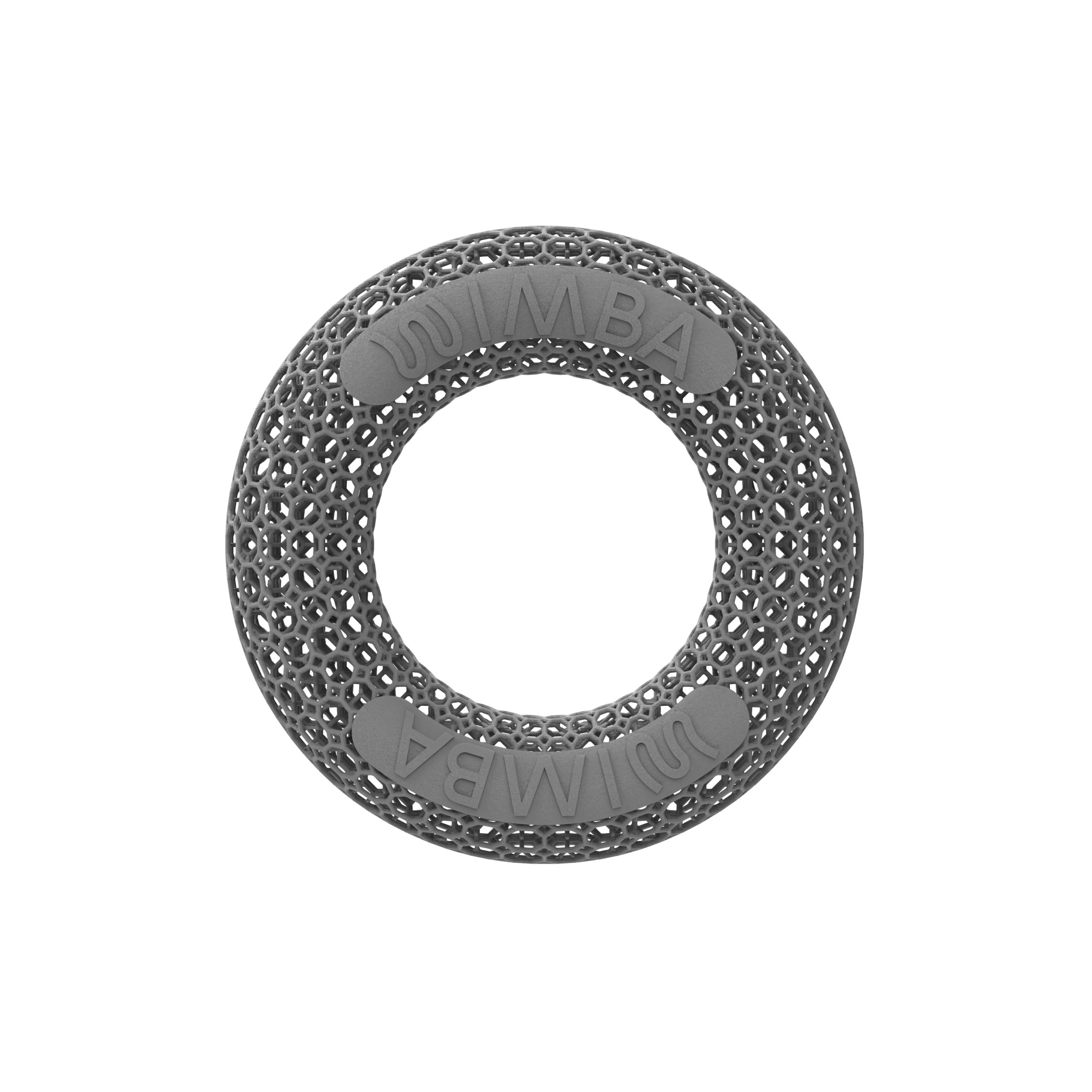
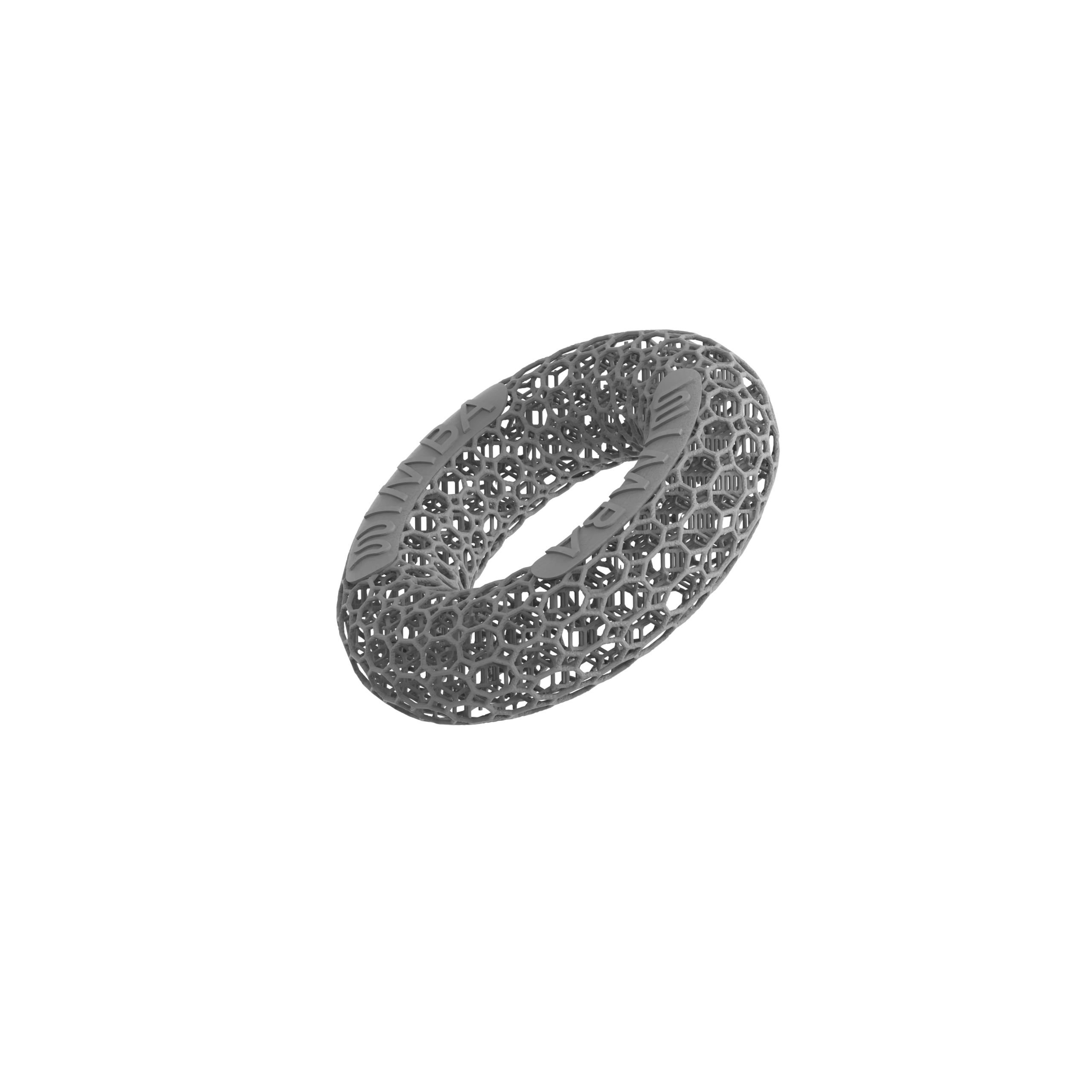
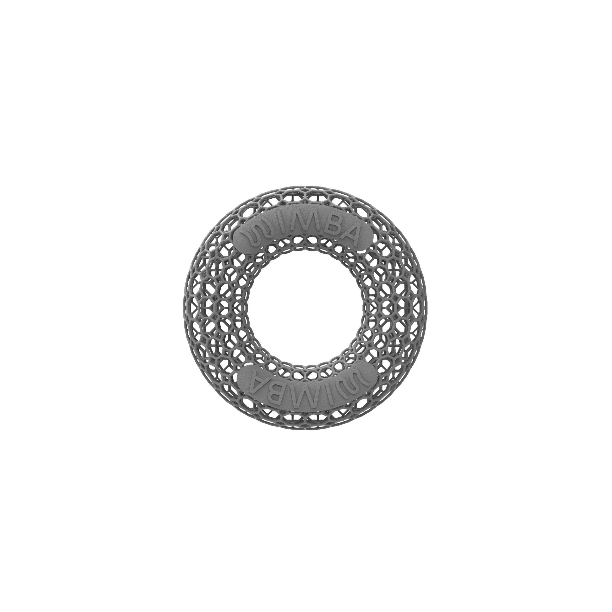
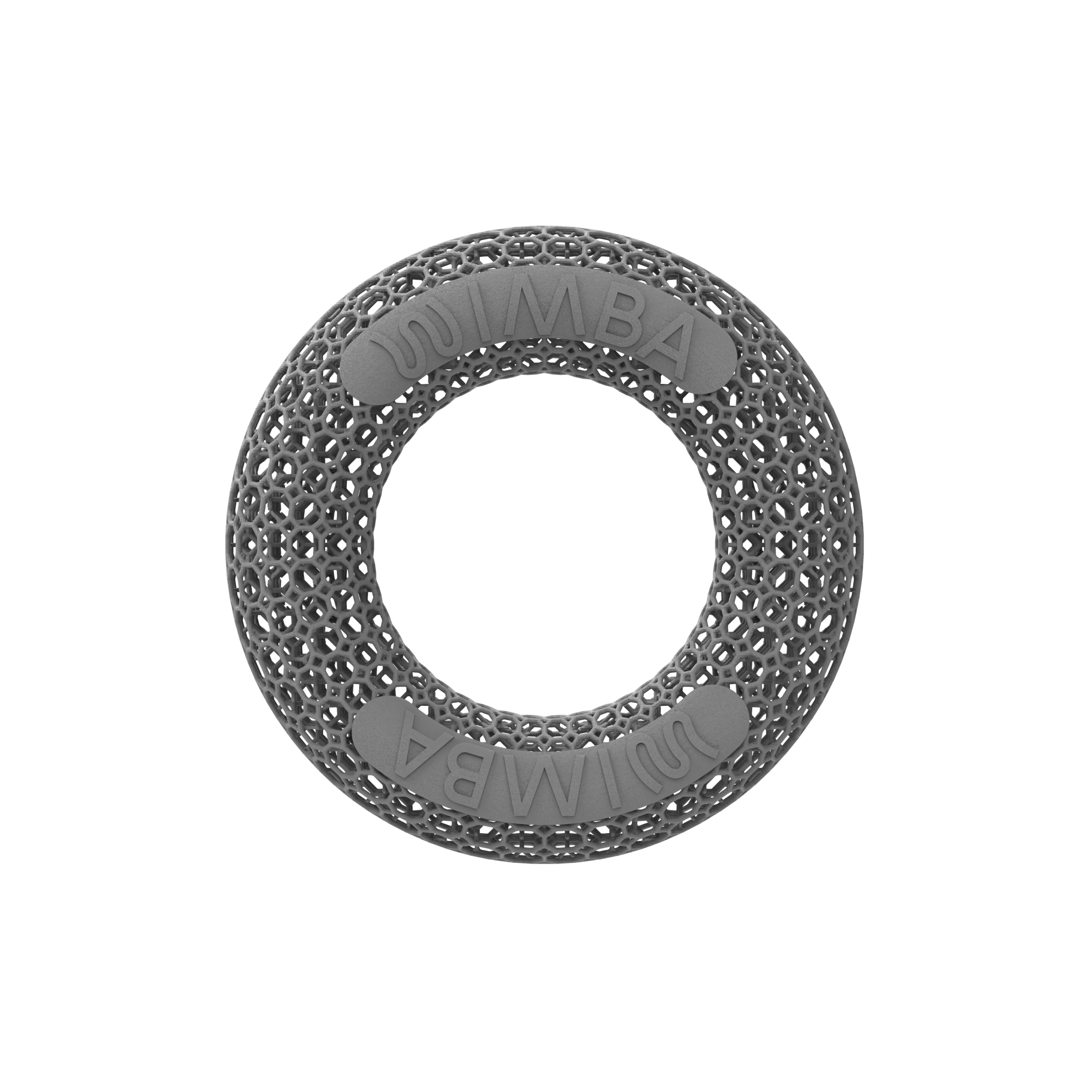
- Donut: ring-shaped clearance around focal or approximately round wounds, calluses, pressure sores, and prominences
- Dome: uniform protective coverage for a selected wound or pressure-sensitive area
- Gradient Dome: a dome with a stiffness gradient for areas surrounded by more delicate tissue
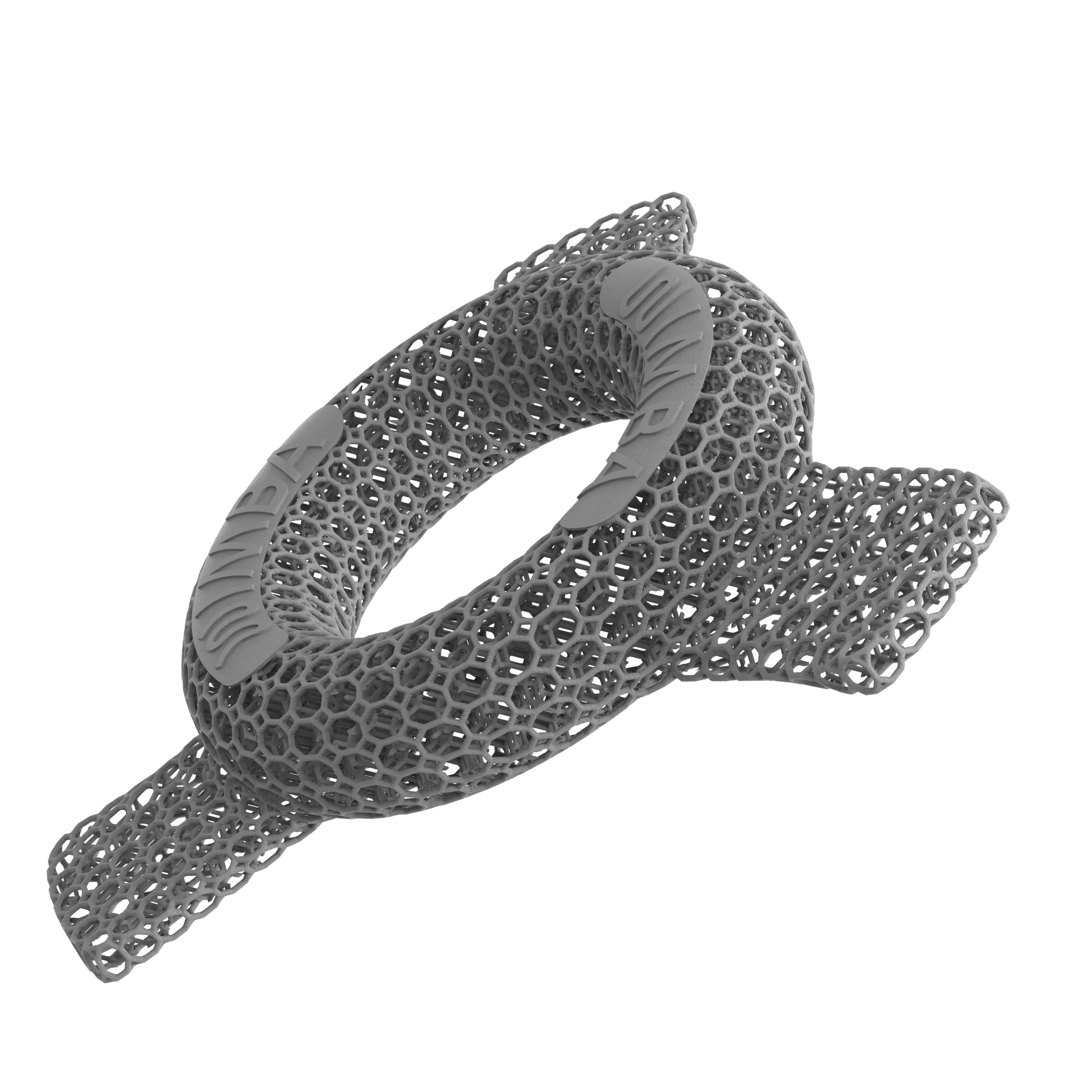
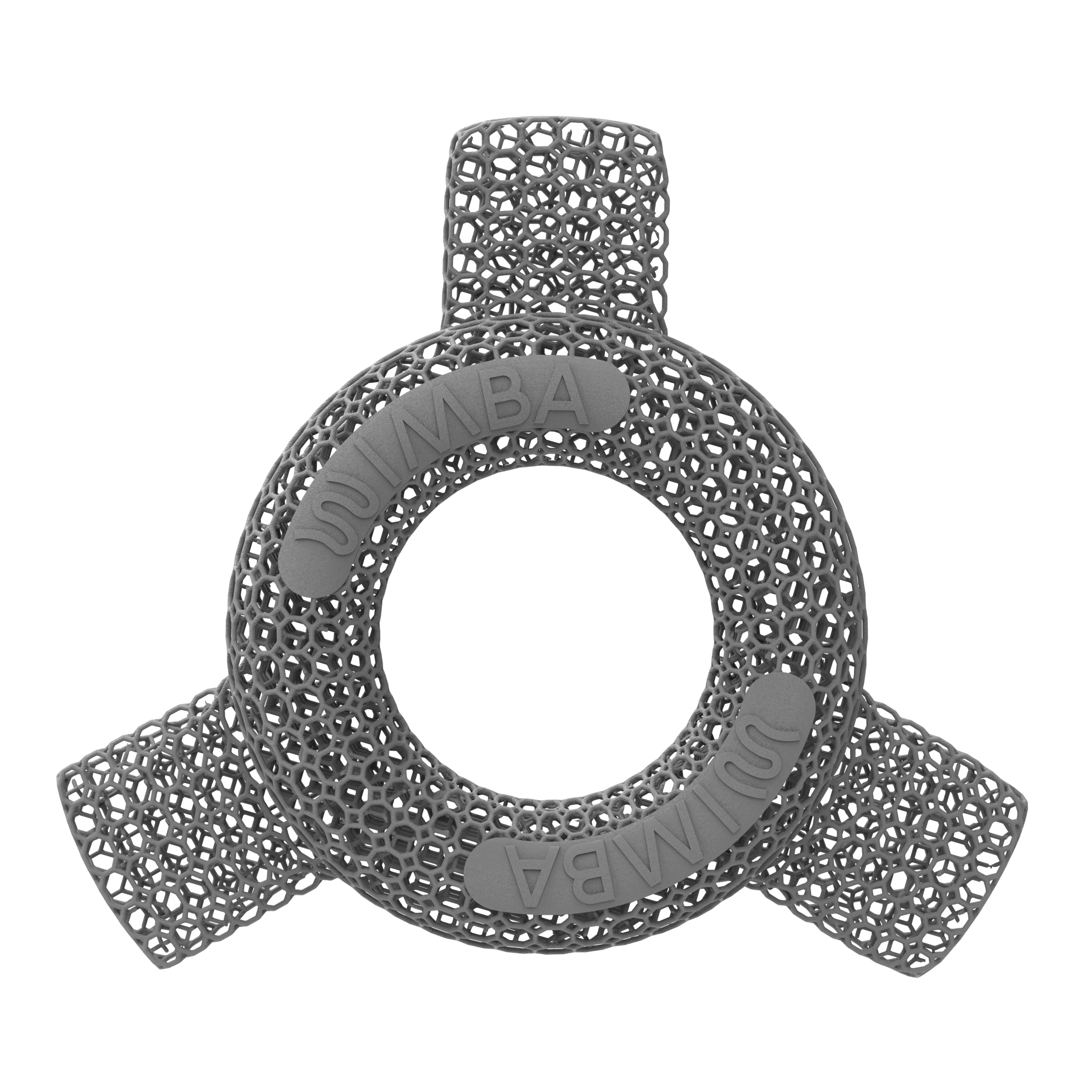
- Donut Wings: additional elements used with proper bandaging to improve retention of the Large Donut
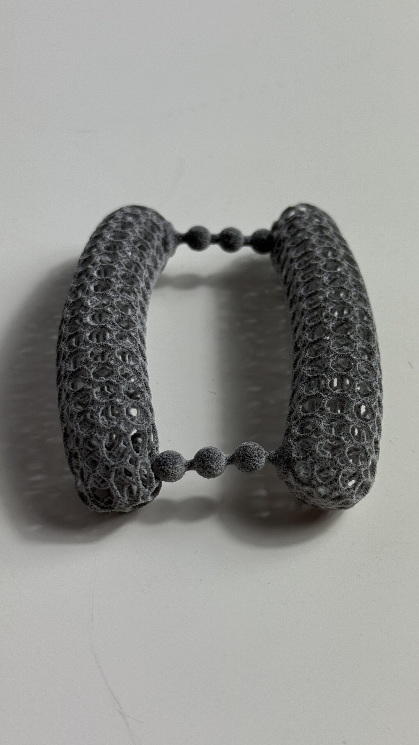
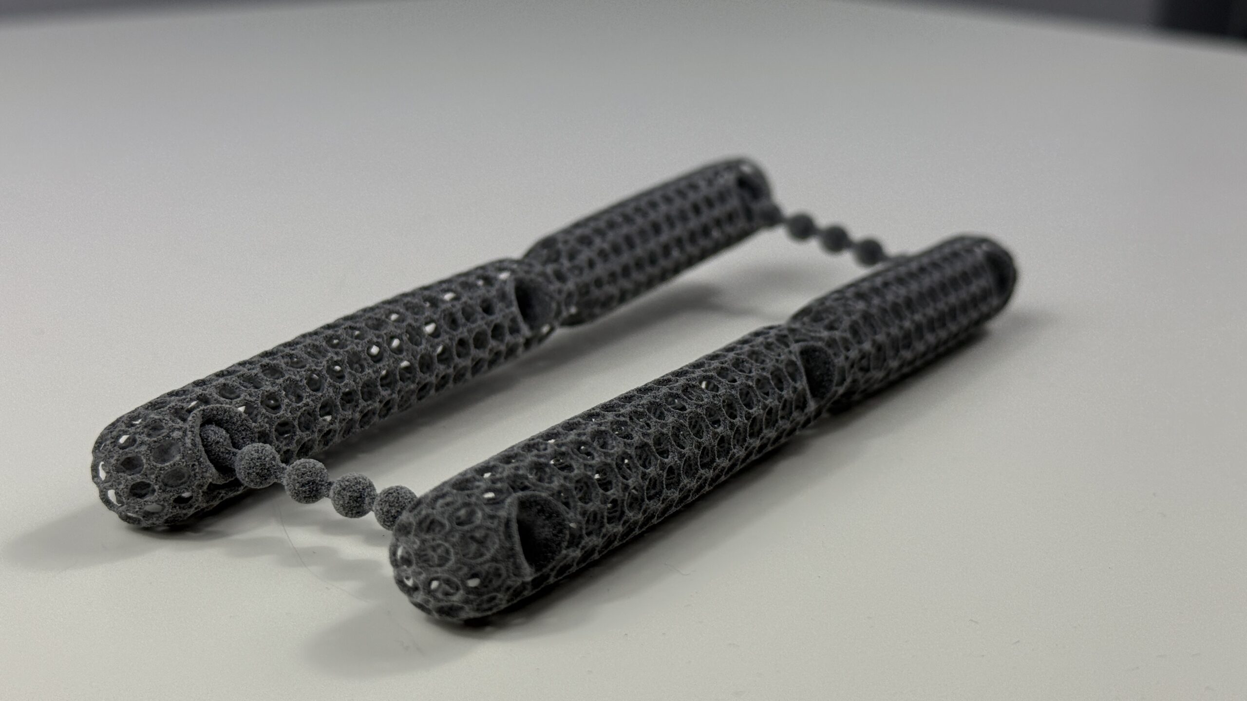
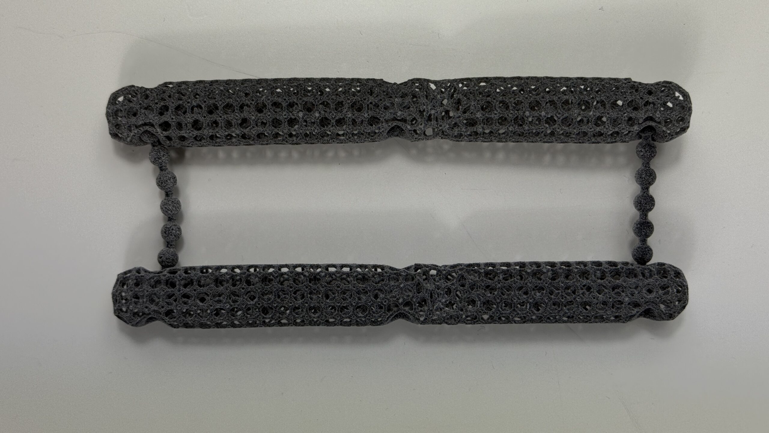
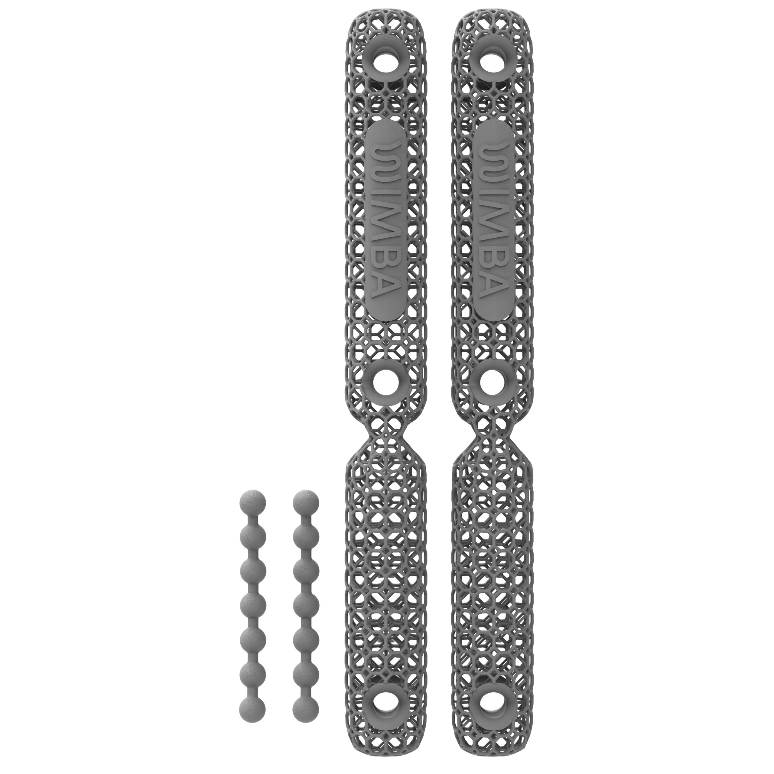
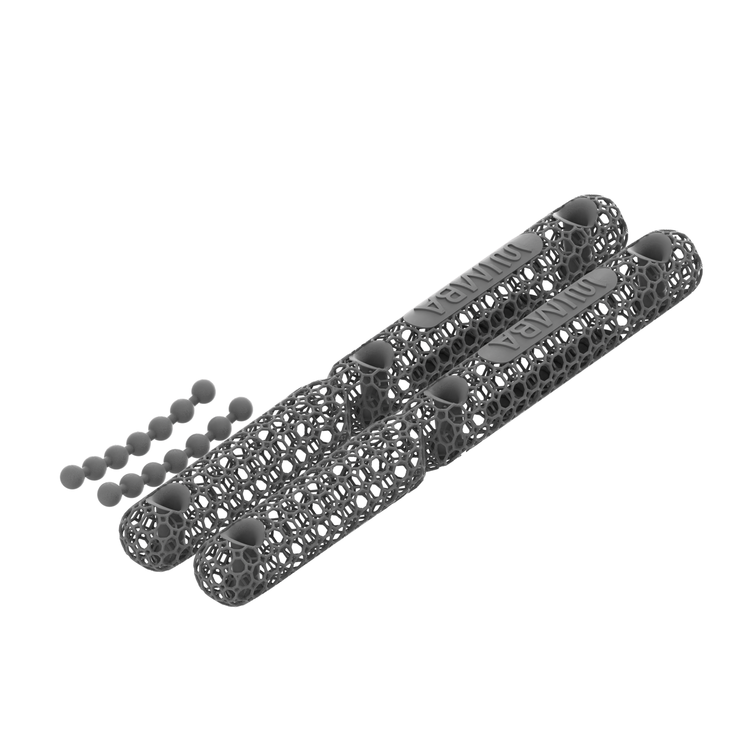
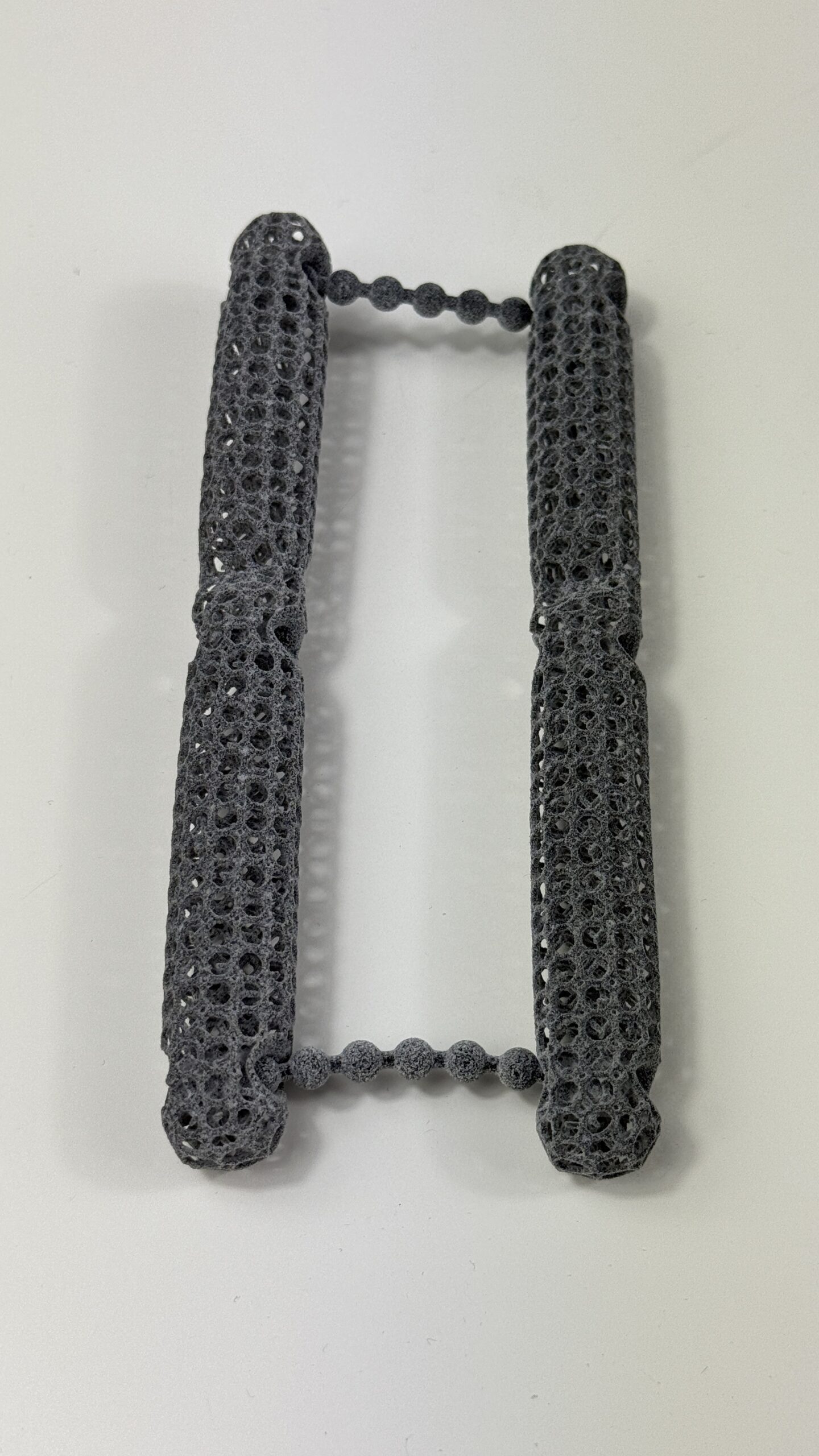
- Care Sticks: trimmable spacer elements for linear wounds, lacerations, tears, or sutured areas
Selection still requires clinical judgement. The product should create the intended clearance without resting on the wound, concentrating force at its edge, impairing circulation, or destabilising the bandage. Care Sticks may be shortened only at the designated narrow connection points in accordance with current product guidance; other components should not be improvised or structurally altered.
WIMBA Wound Care can be hand-cleaned with lukewarm water and surgical or mild pH-neutral soap, air-dried, and reused up to five times under the current instructions. Reusable does not mean sterile or suitable for indefinite use. The clinician should discard a component if cleanliness, integrity, geometry, or safe clinical use is uncertain.
Clinics currently order through the WIMBA Wound Care order page or an authorised regional distributor. The WimbaAPP workflow described for custom orthoses should not be presented as the ordering route for this ready-to-use product line.
What a Protective Spacer Cannot Replace
A spacer is not a wound-contact dressing, antimicrobial treatment, debridement method, drainage system, or diagnostic test. It does not close a dehisced incision, restore perfusion, remove necrotic tissue, treat osteomyelitis, control an unstable joint, or compensate for a wet, slipping, contaminated, or excessively tight bandage.
Complex wounds may require lavage, debridement, culture and targeted antimicrobial therapy, surgical closure, reconstruction, negative-pressure wound therapy, or second-intention management. Canine NPWT research shows why modalities cannot be treated as interchangeable: an experimental study found earlier smooth granulation with NPWT but less contraction and epithelialisation during prolonged use, while a prospective clinical series of 45 dogs described NPWT as one stage before closure or second-intention healing.[9][10]
Dressing evidence is also product-specific. A 2024 controlled study in six Beagles found different healing outcomes among nitric oxide, hydrocolloid, and control dressings, but it did not evaluate a WIMBA spacer.[11] The appropriate primary layer and treatment schedule remain patient-specific decisions within veterinary wound management.
Application, Monitoring, and Stop Criteria
Before application, photograph and measure the wound, inspect surrounding skin, and document perfusion, sensation, pain, swelling, and function. Apply the prescribed primary dressing, position the selected spacer over that covered site, and secure the complete construct using sound bandaging technique. Confirm that the device remains stable in the positions and controlled activities expected for that patient.
Recheck intervals should reflect wound type, exudate, infection risk, bandage design, location, patient behaviour, mobility, and owner competence. Do not assume that a comfortable dog or normal-looking exposed toes proves that all covered tissue is healthy. Cast research found that most soft-tissue injuries were identified by veterinary professionals and that injury could occur at any point during the casting period.[4]
Owners need written instructions to keep the bandage clean and dry, prevent chewing, follow activity and positioning guidance, and attend scheduled changes. Earlier examination is warranted for slippage, wetness, odour, discharge, increasing pain, agitation, swelling, cold or discoloured exposed digits, loss of function, fever, lethargy, or any visible deterioration.
At each change, remove the construct and inspect the entire covered area. Compare the wound with baseline measurements, evaluate every contact edge, and decide whether the spacer still achieves its objective. Effective canine wound care changes course when the clinical response differs from the plan.
A Practical Clinic Workflow
- Diagnose and document the wound, patient factors, and any pressure or contact source.
- Define a measurable objective, such as clearance over a focal site or protection of a linear incision.
- Select the primary dressing and complete treatment plan before choosing an external spacer.
- Choose the appropriate Donut, Dome, Gradient Dome, Donut Wings, or Care Sticks configuration.
- Apply the spacer over the covered wound and stabilise it without creating focal pressure or constriction.
- Inspect the patient in relevant positions and controlled movement, then record the finished construct.
- Provide written home-care, repositioning, activity, bandage-protection, and stop instructions.
- Reassess the wound and all covered tissue at every change; continue only while benefit exceeds risk.
This process makes pressure offloading for dogs one controlled element of care rather than an automatic response to every slow-healing wound. It also creates records that show whether the intervention is associated with improved clearance, stability, tissue appearance, and wound progression.
Evidence Boundaries and Responsible Communication
The peer-reviewed studies cited here support the importance of pressure, positioning, bandage design, wound assessment, and modality-specific treatment. They do not constitute controlled clinical evidence that WIMBA products prevent infection, eliminate dehiscence, or accelerate healing in every patient.
Clinics using wound care spacers should document outcomes consistently: wound dimensions, tissue appearance, exudate, pain, surrounding skin, spacer position, bandage changes, concurrent treatment, and adverse events. Aggregated prospective data would help determine which wounds benefit, how often displacement or new pressure points occur, and whether healing differs from comparable cases managed without a spacer. Until such studies exist, clinical conclusions should remain appropriately cautious.
WIMBA patient stories can illustrate clinical use, but they are observational examples involving concurrent treatment. They cannot establish causation or an expected healing time. Marketing should therefore describe Wound Care spacers as protective adjuncts designed to create clearance, distribute external forces, and support airflow when used over a primary dressing.
Avoid claims that the product heals infection, guarantees wound closure, replaces repositioning, or prevents all pressure injury. The most defensible message is that WIMBA Wound Care gives veterinary teams an additional way to protect selected covered wounds from direct mechanical stress within a monitored treatment plan.
Frequently Asked Questions About Wound Complications in Dogs
What causes wound complications in dogs?
Possible causes include infection, devitalised tissue, foreign material, poor perfusion, systemic disease, malnutrition, self-trauma, excessive movement, moisture imbalance, pressure, friction, shear, or an unsuitable bandage. A veterinary examination is needed because similar wound changes can have different causes.
Are WIMBA wound care spacers custom-made?
No. The current WIMBA Wound Care range consists of standard 3D-printed Donut, Dome, Gradient Dome, Donut Wings, and Care Sticks components in defined forms and sizes. The clinic selects the appropriate option for the wound and bandage plan; no patient scan or custom manufacturing submission is required.
Can a WIMBA spacer be placed directly on a wound?
No. A suitable primary wound dressing must cover the wound first. The spacer is positioned over that protective layer and secured within the outer bandage construct to create clearance and reduce direct external pressure.
Which WIMBA Wound Care component should a clinic choose?
Selection depends on wound shape, size, location, surrounding tissue, pressure source, patient position, and bandage retention. Donuts suit focal or approximately round sites, Domes provide protective coverage, Gradient Domes add a stiffness gradient, Donut Wings assist retention of the Large Donut, and Care Sticks suit linear sites.
Do wound care spacers replace primary dressings or wound treatment?
No. They are external protective adjuncts. Debridement, lavage, infection control, primary dressing selection, surgery, analgesia, repositioning, nutrition, and treatment of systemic disease remain separate clinical decisions.
How many times can WIMBA Wound Care be reused?
Current instructions allow hand-cleaning with lukewarm water and surgical or mild pH-neutral soap, air-drying, and reuse up to five times. Reuse is appropriate only when the component remains clean, intact, correctly shaped, and clinically suitable.
How can a veterinary clinic order WIMBA Wound Care?
Clinics can use the current WIMBA Wound Care order page or contact an authorised distributor in an available region. These ready-to-use spacers do not require the WimbaAPP scanning and custom-order workflow used for patient-specific WIMBA orthoses.
Medical and Product Disclaimer: This material is intended for veterinary professional education and general product information. It does not replace patient-specific examination, diagnosis, wound treatment, bandaging competence, prescribing, surgery, or emergency care. Product forms, sizes, instructions, availability, cleaning limits, and ordering routes may change. Follow the current manufacturer instructions and applicable professional regulations. No healing time, infection outcome, or clinical result is guaranteed.
Scientific References
- Lux CN. Wound healing in animals: a review of physiology and clinical evaluation. Vet Dermatol. 2022;33(1):91–e27. doi:10.1111/vde.13032
- Caraty J, De Vreught L, Cachon T, Moissonnier P, Bongartz A, Viguier E, Carozzo C. Comparison of the different supports used in veterinary medicine for pressure sore prevention. J Small Anim Pract. 2019;60(10):623–630. doi:10.1111/jsap.13061
- Tambella AM, Attili AR, Dini F, Palumbo Piccionello A, Vullo C, Serri E, Scrollavezza P, Dupré G. Autologous platelet gel to treat chronic decubital ulcers: a randomized, blind controlled clinical trial in dogs. Vet Surg. 2014;43(6):726–733. doi:10.1111/j.1532-950X.2014.12148.x
- Meeson RL, Davidson C, Arthurs GI. Soft-tissue injuries associated with cast application for distal limb orthopaedic conditions: a retrospective study of sixty dogs and cats. Vet Comp Orthop Traumatol. 2011;24(2):126–131. doi:10.3415/VCOT-10-03-0033
- Anderson DM, White RAS. Ischemic bandage injuries: a case series and review of the literature. Vet Surg. 2000;29(6):488–498. doi:10.1053/jvet.2000.17847
- Thibodaux B, Eginton K, Gordon-Evans W. Sub-bandage pressure changes in canine hindlimbs after re-application of custom rigid fiberglass splints. BMC Vet Res. 2024;20:574. doi:10.1186/s12917-024-04437-6
- Iodence AE, Olsen AM, McGilvray KC, Duncan CG, Duerr FM. Use of pressure mapping for quantitative analysis of pressure points induced by external coaptation of the distal portion of the pelvic limb of dogs. Am J Vet Res. 2018;79(3):317–323. doi:10.2460/ajvr.79.3.317
- Swaim SF, Vaughn DM, Spalding PJ, Riddell KP, McGuire JA. Evaluation of the dermal effects of cast padding in coaptation casts on dogs. Am J Vet Res. 1992;53(7):1266–1272. doi:10.2460/ajvr.1992.53.7.1266
- Demaria M, Stanley BJ, Hauptman JG, Steficek BA, Fritz MC, Ryan JM, Lam NA, Moore TW, Hadley HS. Effects of negative pressure wound therapy on healing of open wounds in dogs. Vet Surg. 2011;40(6):658–669. doi:10.1111/j.1532-950X.2011.00849.x
- Pitt KA, Stanley BJ. Negative pressure wound therapy: experience in 45 dogs. Vet Surg. 2014;43(4):380–387. doi:10.1111/j.1532-950X.2014.12155.x
- Rodriguez-Diaz JM, Wallace ML, Emond SA, McNabney D, Hodges KM, Howerth EW. Effect of hydrocolloid-nitric oxide wound dressings on wound healing in dogs. Vet Surg. 2024;53(8):1351–1365. doi:10.1111/vsu.14149
Related reading: Wound Care Spacers for Complex Orthopedic Cases: A Veterinary Guide · Instruction for Use WIMBA Wound Care