 Skip to content

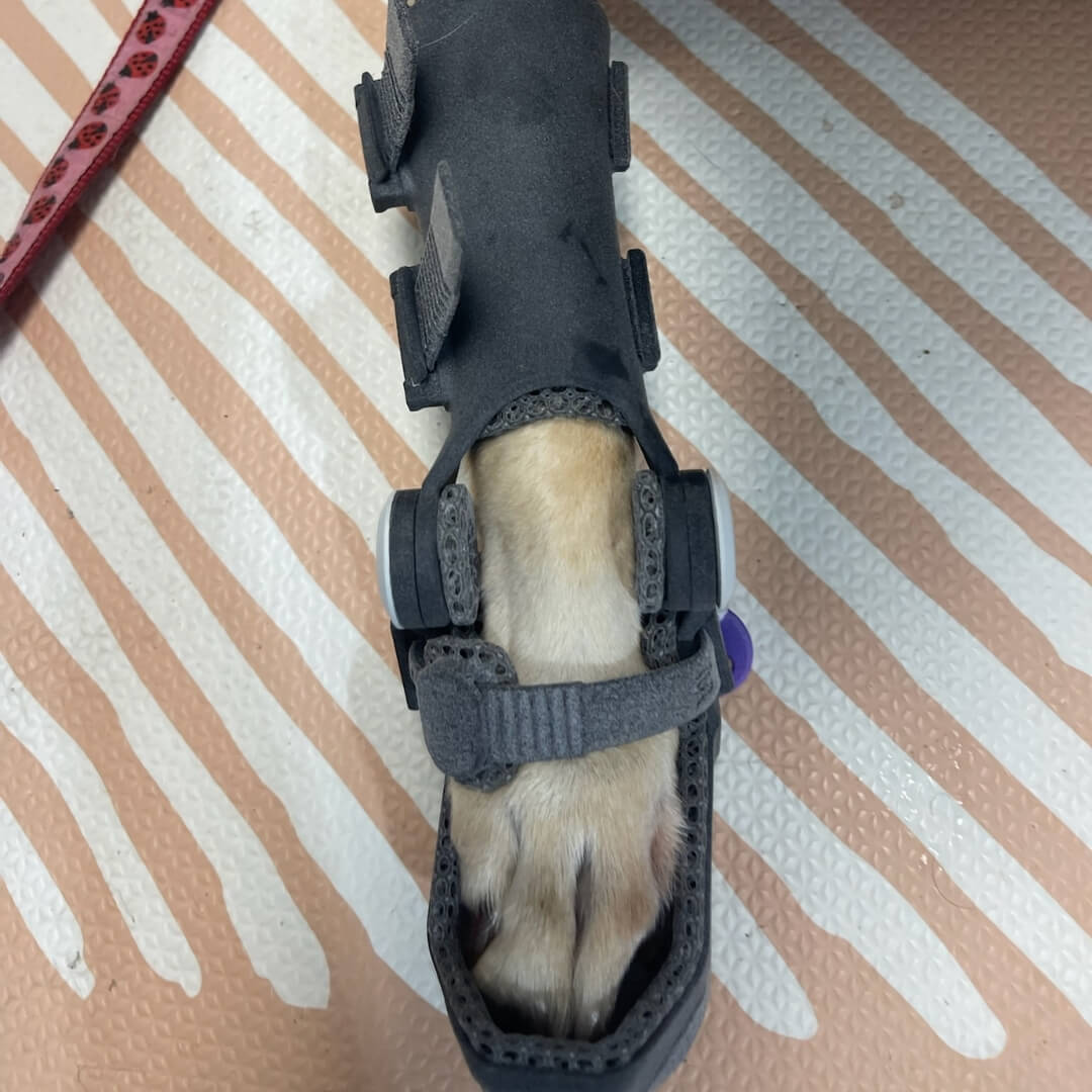
3D printed dog orthotics replace physical casting and manual shell fabrication with a digital design and manufacturing workflow. For veterinary teams, the practical differences extend beyond the printer itself. Data collection, case review, material selection, repeatability, fitting, adjustment, and access to the service all influence the final result.
This guide compares 3D printed dog orthotics with traditional cast-based orthotics without assuming that one manufacturing method guarantees a better clinical outcome. It explains how WIMBA produces custom dog orthotics, where digital workflows offer practical advantages, and why diagnosis, fit, monitoring, and rehabilitation remain essential with every type of brace.
3D Printed Dog Orthotics vs Traditional Manufacturing
The central difference is how patient anatomy becomes a finished device. A cast-based workflow creates a physical negative impression and positive mould before a thermoplastic shell is shaped by hand. 3D printed dog orthotics begin with structured digital information, continue through computer-aided design, and are manufactured directly from the approved digital file. This distinction shapes how digital veterinary orthotics are ordered, reproduced, and revised.
Both pathways still depend on clinical reasoning. Neither casting nor scanning identifies the diagnosis, selects the correct joint, defines the required plane of support, or determines the rehabilitation programme. The manufacturing route can improve or complicate the workflow, but it cannot compensate for unsuitable case selection.
| Category | Traditional cast-based workflow | Digital 3D-printing workflow |
|---|---|---|
| Patient data | Physical impression of the limb | Guided measurements and photographs or a digital limb scan |
| Design | Positive mould and manual shaping | Computer-aided, patient-specific design |
| Manufacturing | Thermoplastic formed over the mould | Additive manufacturing from a digital file |
| Repeatability | Relies on the retained mould and manual recreation | Design data can be stored, reviewed, and revised |
| Local adjustment | Some thermoplastics can be heat-adjusted by trained personnel | Structural parts should not be heat-shaped or modified in clinic |
| Remote access | Often requires transfer of a physical cast or specialist appointment | Patient data can be submitted digitally by an authorised veterinary clinic |
How Traditional Cast-Based Orthotics Are Made
Traditional fabrication generally starts with a plaster or fibreglass impression of the affected limb in a prescribed position. That negative impression is used to create a positive model. A thermoplastic sheet is then heated, formed over the model, trimmed, finished, padded, and fitted with hinges, straps, or closures as required.
The process is established and can be highly effective in experienced hands. Some thermoplastic components can also be locally adjusted after fitting. However, the final geometry depends on several manual stages, including patient positioning, impression technique, correction of the positive mould, material forming, trimming, and assembly.
Potential strengths of traditional fabrication:
- Long-established clinical and fabrication methods
- Direct physical impression of the limb
- Possibility of selected heat adjustments by an appropriately trained professional
- Useful where an experienced veterinary orthotist is locally available
Potential limitations: multiple manual stages, patient tolerance of casting, shipment or storage of physical models, and dependence on the consistency of impression-taking and fabrication.
How 3D Printed Dog Orthotics Are Made
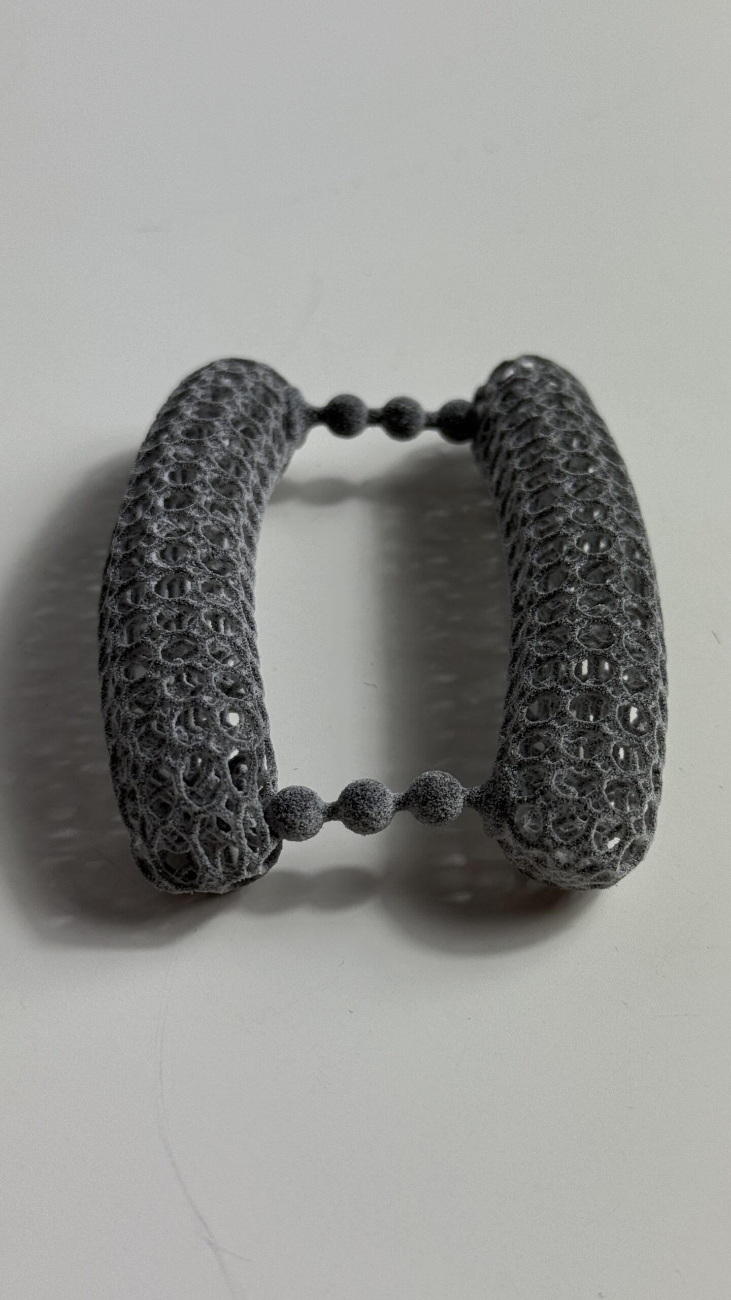
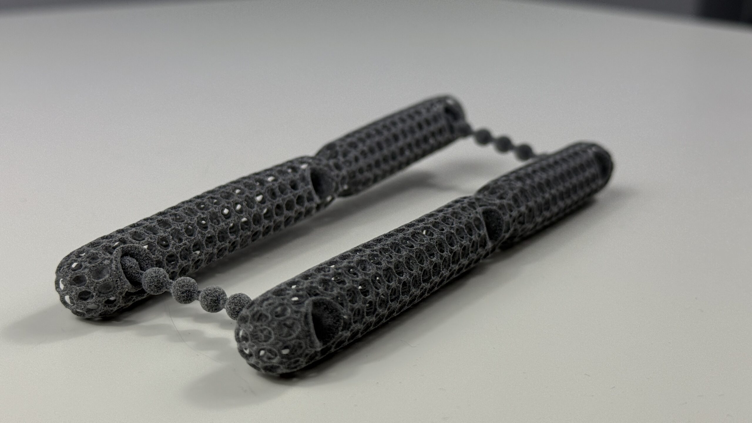
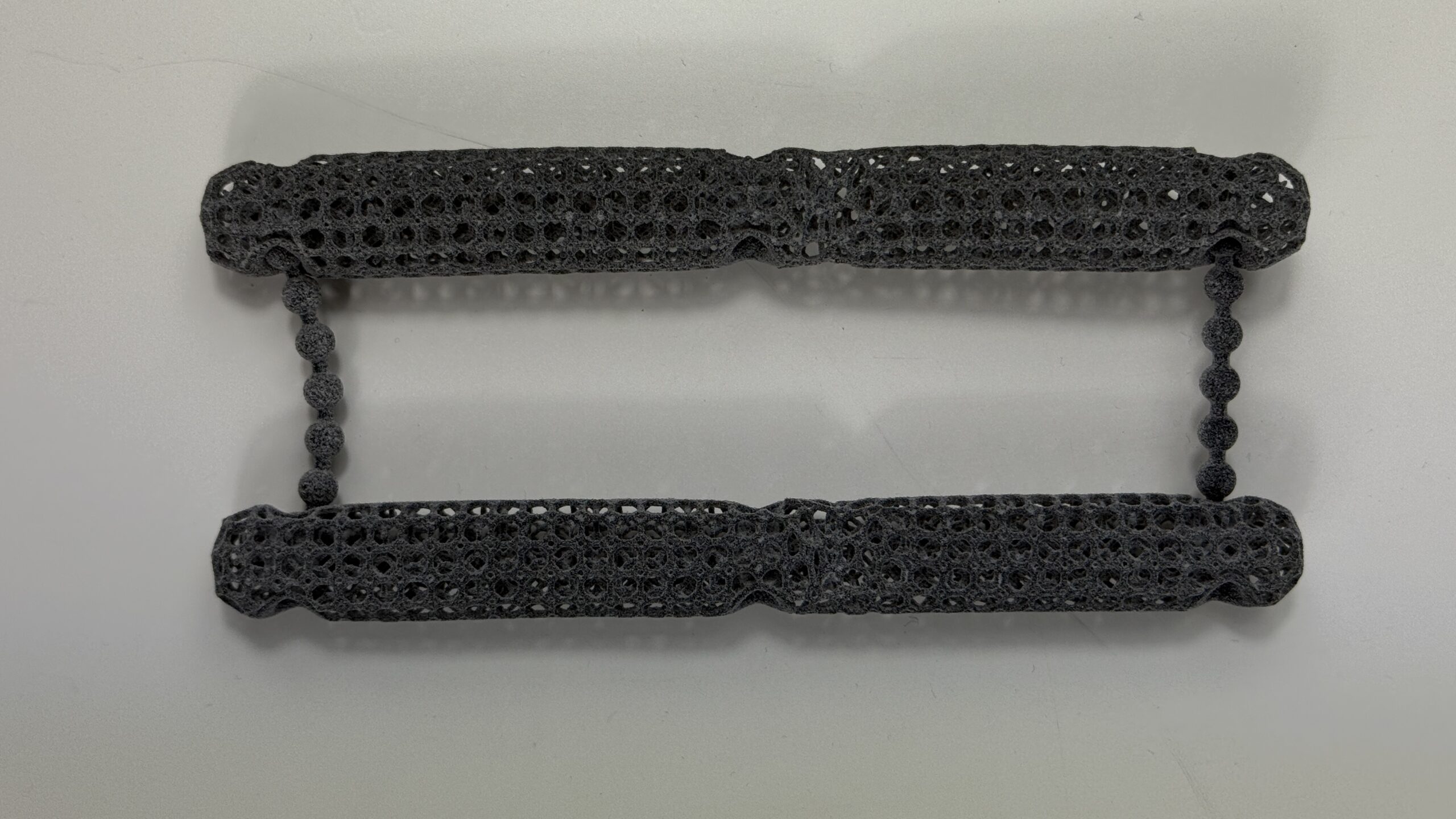
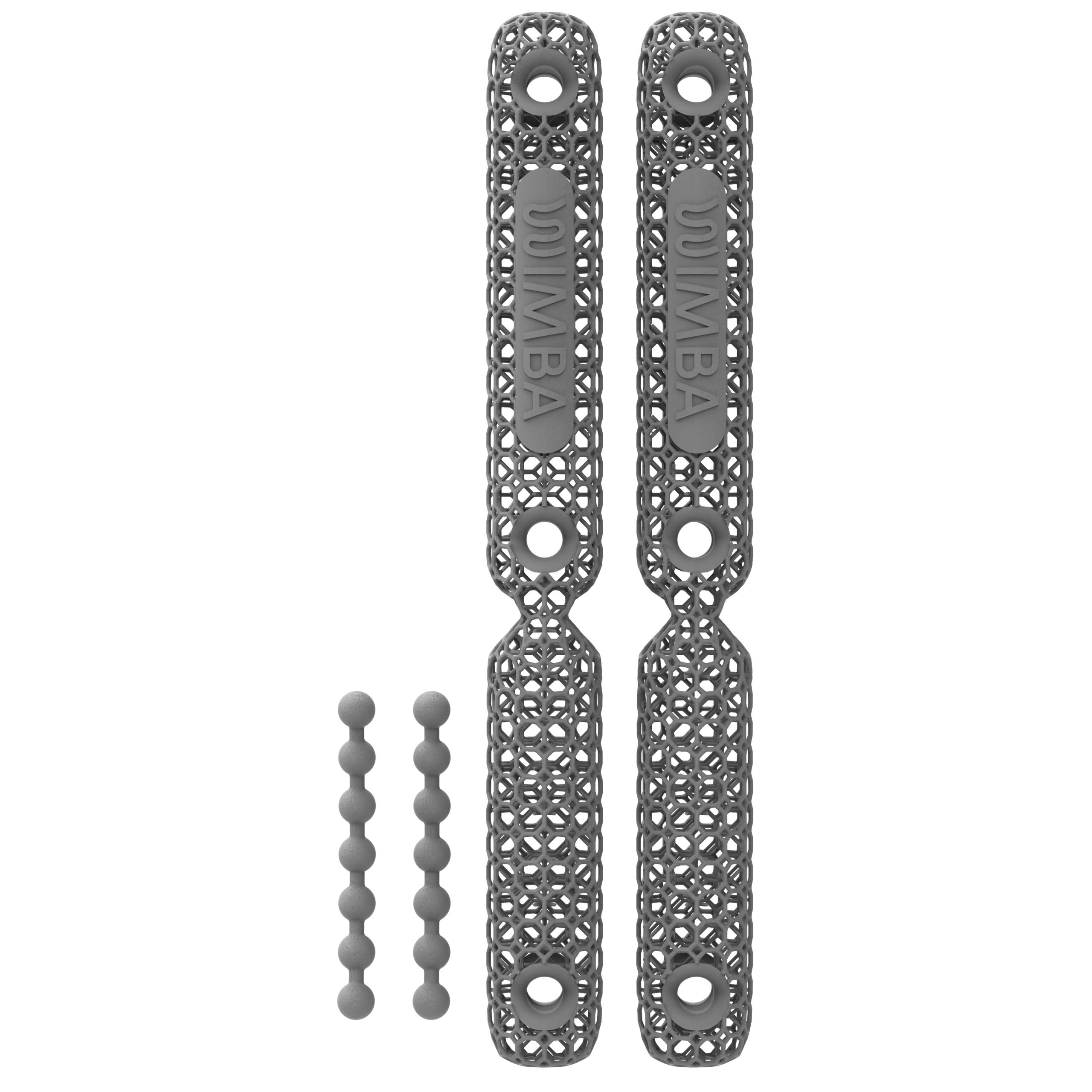
3D printed dog orthotics use patient data to create a digital representation of the required anatomy. The designer uses that information together with the prescription to define the frame, joint position, clearances, closures, range-of-motion components, and other functional features. The approved parts are then produced layer by layer and assembled into the finished device.
The quality of digital veterinary orthotics still depends on the input. Inaccurate measurements, blurred photographs, moving anatomy, poor lighting, an incorrectly positioned joint, or incomplete clinical information can all affect design. Digital manufacturing reduces some manual fabrication variability, but it does not make poor patient data harmless.
WIMBA Go: Photographs and Guided Measurements
WIMBA Go provides a streamlined route to selected carpal and tarsal devices. The veterinary clinic submits the required weight-bearing photographs and manual measurements through WimbaAPP. A physical cast and WimbaSCAN are not required. This pathway allows eligible 3D printed dog orthotics to be ordered without a Pro scanning kit.
Go is not simply a lower-data version of every possible device. It is intended for cases that fall within current Go indications and geometry requirements. Complex, multiplanar, markedly deformed, or otherwise atypical cases should be reviewed before the clinic assumes that the measurement-based pathway is sufficient.
WIMBA Pro: WimbaSCAN and Manual Measurements
WIMBA Pro is the scan-based pathway for more complex carpal, tarsal, and stifle devices. WimbaSCAN uses a high-resolution smartphone video recorded around the prepared limb with WimbaBOX components, including a calibration mat and patterned sleeve or wrap. The recording is processed into a three-dimensional model and combined with required manual measurements.
The dog should remain appropriately positioned while the complete limb and calibration area stay visible. WIMBA’s workflow is designed around an awake, standing or safely supported patient. Sedation is not an inherent step in ordering 3D printed dog orthotics. If positioning cannot be achieved safely, the clinic should discuss the case with WIMBA rather than treating sedation as a routine scanning requirement.
Fit and Precision Depend on the Entire Workflow
The phrase “digitally made” should not be treated as a guarantee of fit. With traditional cast-based orthotics, errors may arise during impression-taking, mould correction, or manual shaping. With 3D printed dog orthotics, errors may arise during photography, measurement, scanning, reconstruction, prescription, digital design, or fitting.
The advantage of a digital process is traceability. Measurements, images, scan files, and design versions can be reviewed without shipping a physical cast. Design parameters can also be applied systematically. The clinical advantage only materialises when the data are accurate and the finished orthosis is fitted and observed on the patient.
In a WIMBA workflow report involving 69 dogs, data from 66 were accepted on the first attempt. Three required recollection because of lighting or insufficient scan and measurement quality. This operational report is useful for understanding the process, but it is not a peer-reviewed comparison of clinical outcomes between 3D printed veterinary orthotics and cast-based devices.
A clinically acceptable fit requires:
- Correct diagnosis and biomechanical objective
- Accurate patient positioning and data collection
- Alignment of hinges with the intended joint region
- No harmful focal pressure from the frame, edges, or closures
- Stability during standing and controlled walking
- A structured acclimatisation and recheck plan
Turnaround and Access with 3D Printed Dog Orthotics
Traditional fabrication may involve an appointment with an orthotist, preparation of the impression, transport of a physical cast, manual fabrication, and a return fitting. The exact timeline varies by provider, geography, device complexity, and whether the first cast is usable.
Data for 3D printed dog orthotics can be uploaded digitally by the veterinary clinic. This removes the need to ship a plaster model and can extend access to clinics without a local casting service. That transferability is a practical advantage of digital veterinary orthotics. It does not remove the need for the dog to attend the clinic for assessment, data collection, fitting, owner training, and follow-up.
For 3D printed dog orthotics, remote manufacturing should therefore be understood as a clinic-to-manufacturer workflow, not direct-to-consumer prescribing. WimbaAPP is intended for verified veterinary professionals. Pro devices additionally require the relevant Provider Pro pathway and WimbaBOX equipment.
Materials Used in 3D Printed Veterinary Orthotics
Traditional orthoses commonly combine formed thermoplastic, foam, straps, closures, and mechanical joints. Their performance depends on material grade, thickness, reinforcement, trim lines, assembly, patient weight, and activity. The category includes many different constructions, so it should not be described as one uniform product.
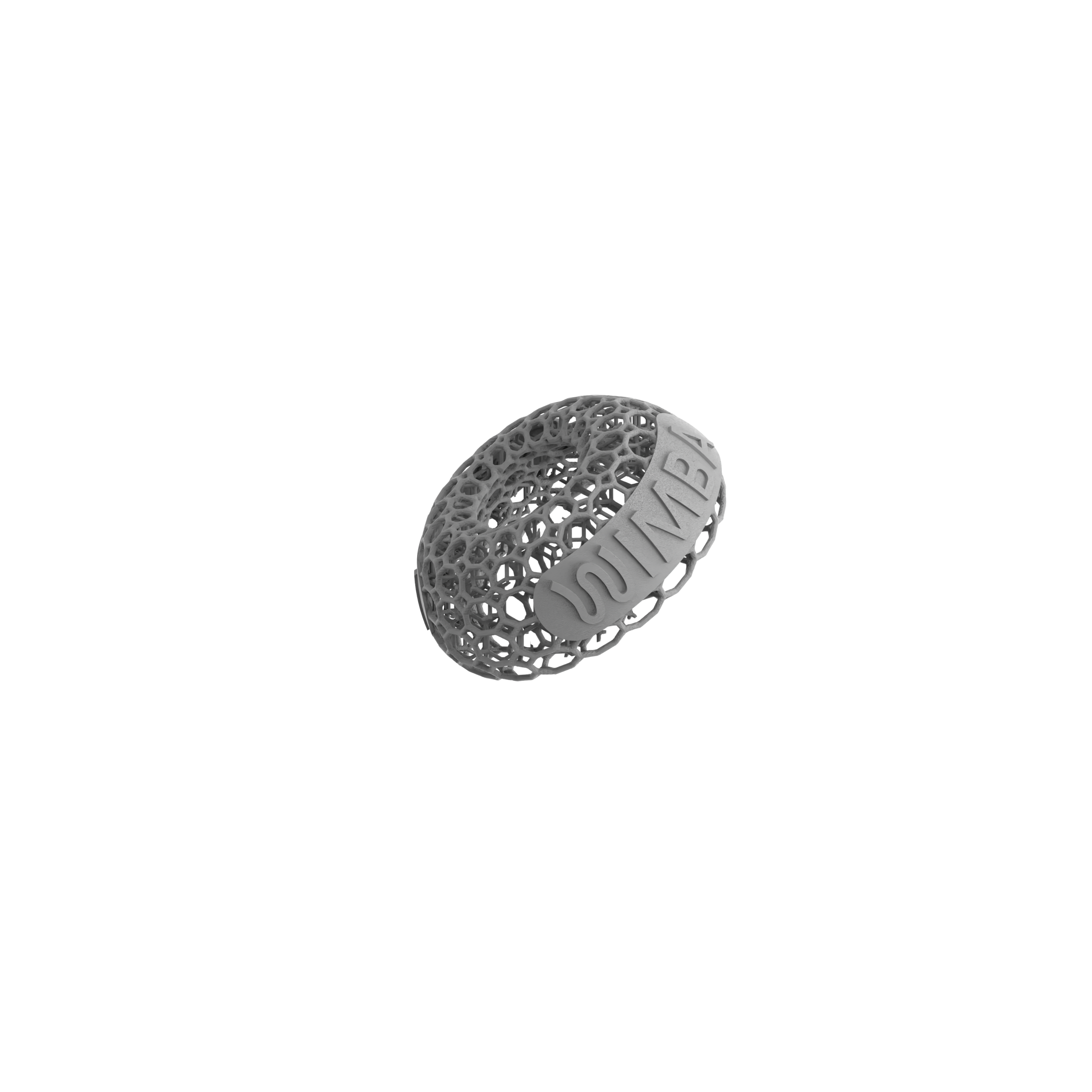
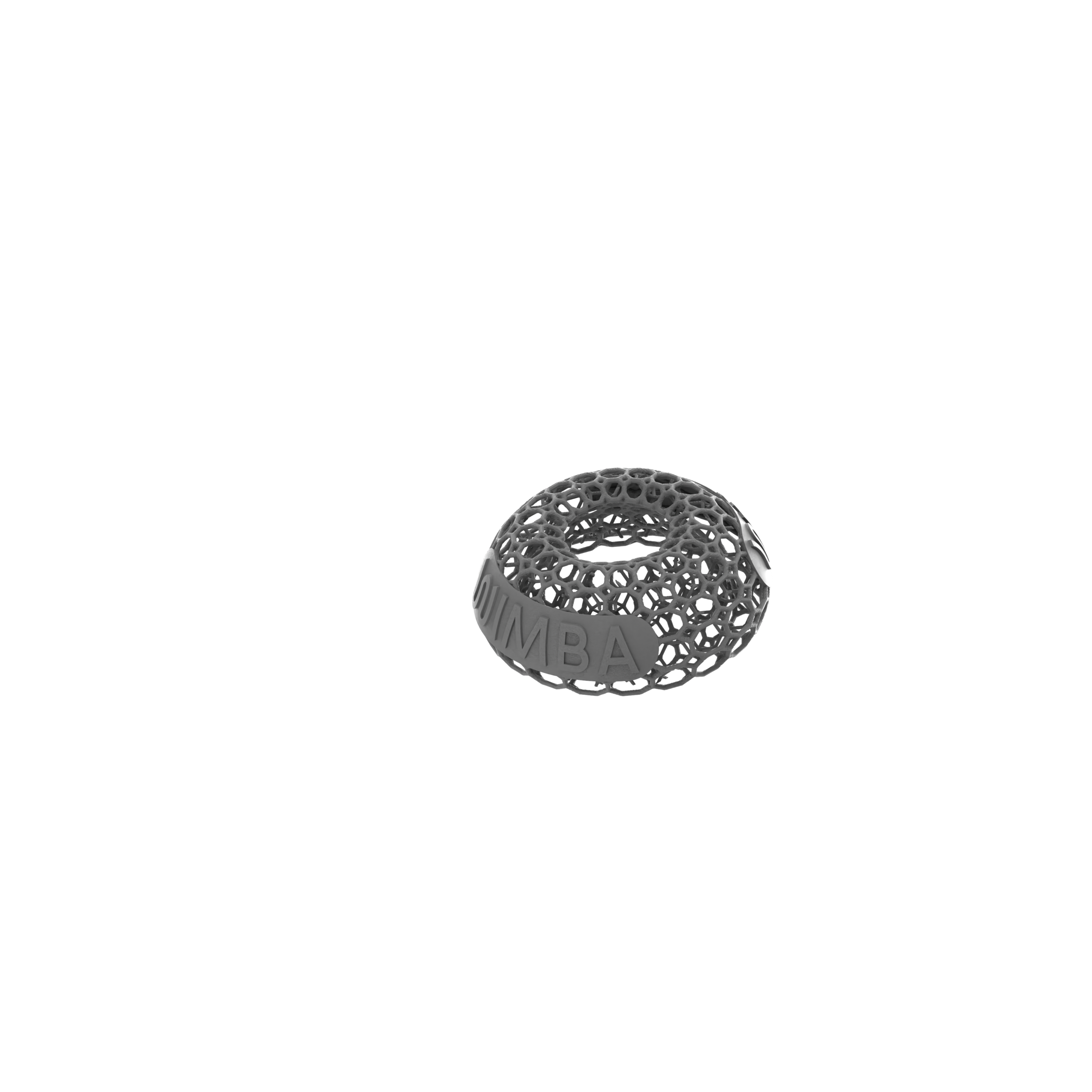
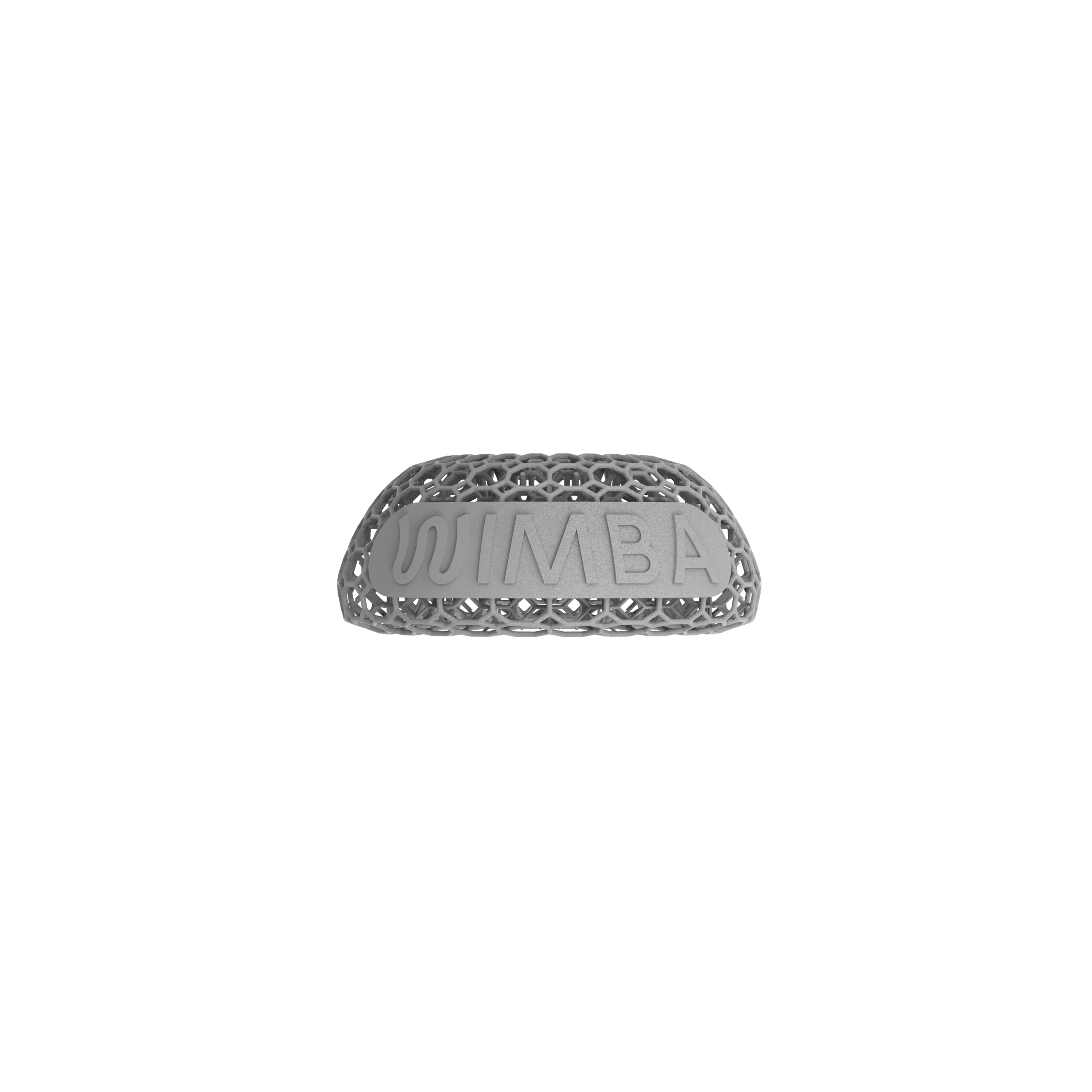
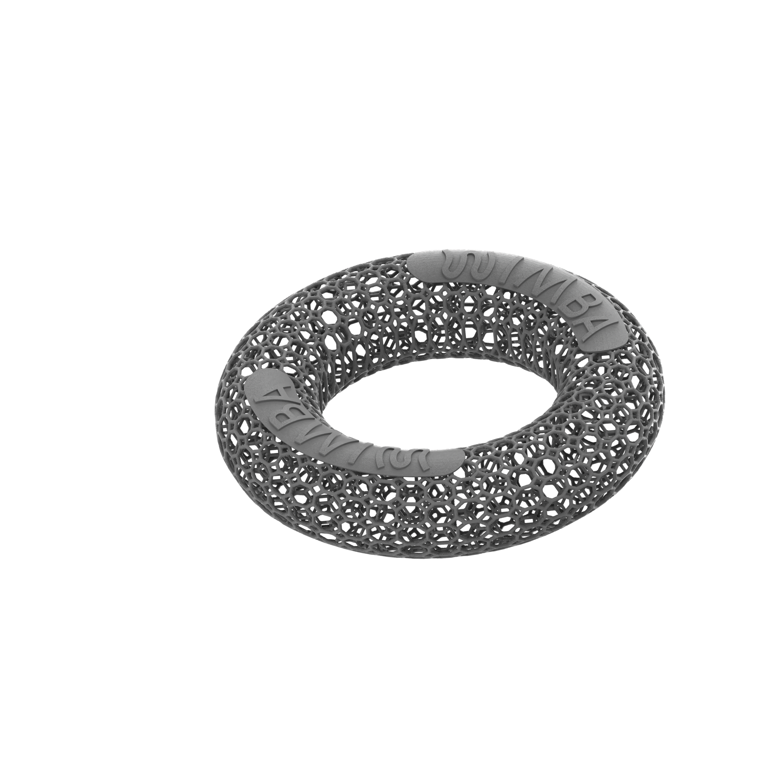
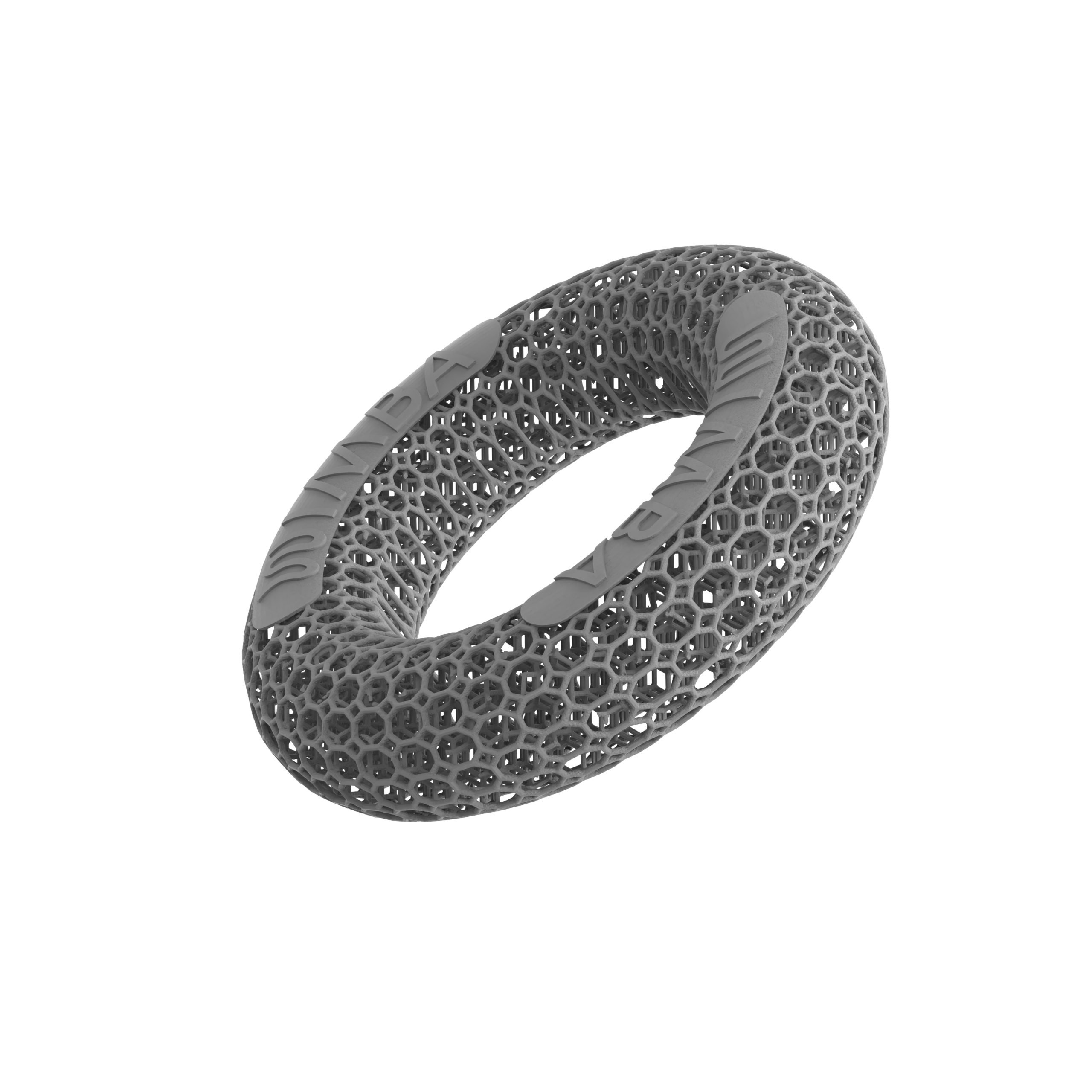
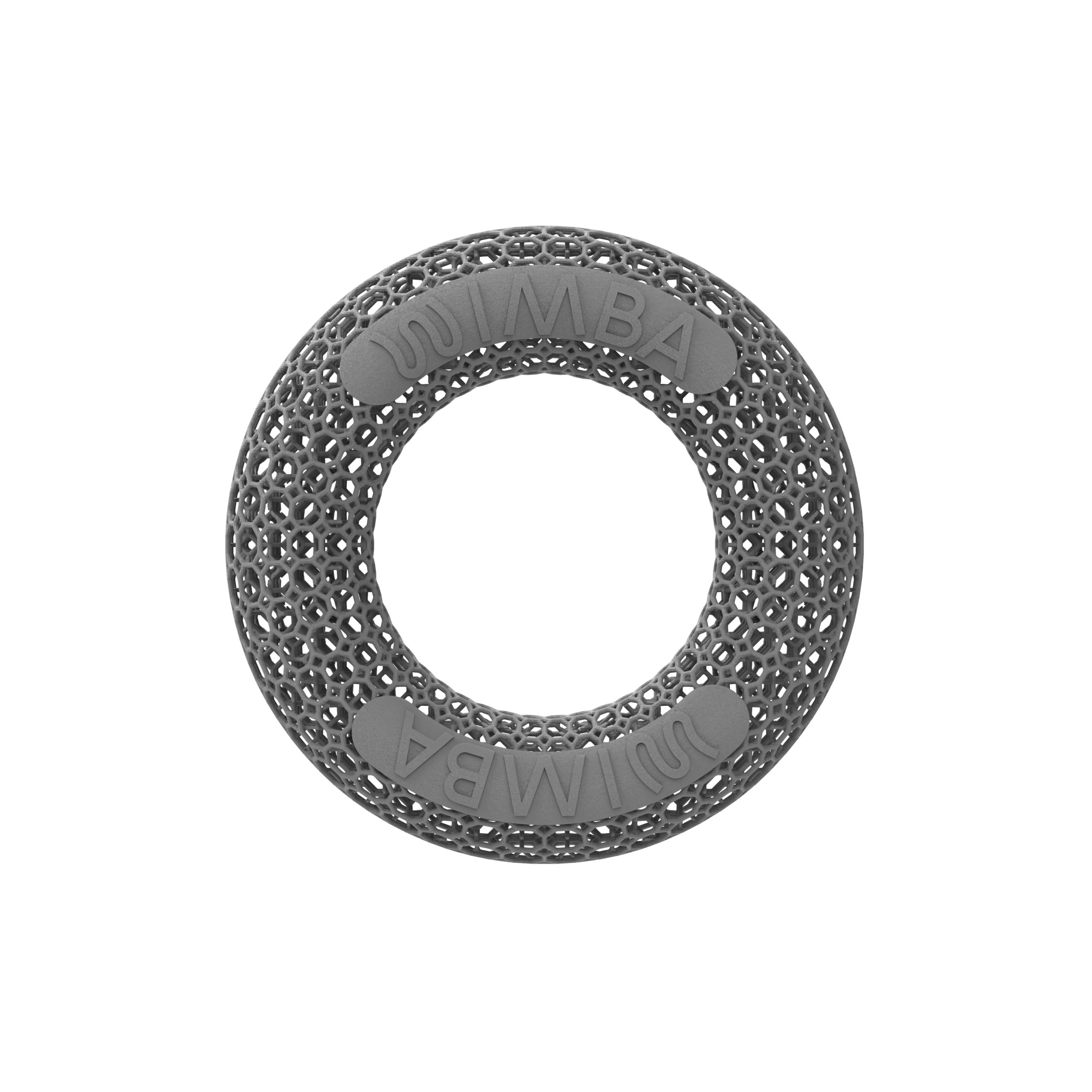
WIMBA manufactures 3D printed veterinary orthotics using HP Multi Jet Fusion. Current WIMBA material information identifies PA11 for the primary frame, PA12 for selected functional parts such as Tokens and hinge caps, and TPU for flexible patient-contact components. Digital design allows material to be distributed according to the intended structure rather than relying only on a uniformly formed sheet.
For 3D printed dog orthotics, this can support lightweight frames, open areas, modular parts, and repeatable component geometry. It does not mean every 3D-printed device is lighter, stronger, or more durable than every traditional one. Valid comparison requires equivalent device purpose, patient size, range of motion, materials, and loading conditions.
Adjustment, Modularity, and Refabrication
One practical strength of some traditional cast-based orthotics is that a trained professional may be able to heat-adjust selected thermoplastic areas. This can help with minor contour changes. Excessive modification can still weaken the device, alter alignment, or introduce new pressure points.
Structural parts of WIMBA 3D printed dog orthotics should not be heated, cut, drilled, or reshaped in clinic. Adjustability instead comes from the device architecture, including straps, replaceable components, inserts, and WIMBA Tokens where applicable. When geometry requires a material change, the digital design can be reviewed and a revised part can be produced.
Stored design data improves traceability and can simplify iteration, but it does not mean old data remain suitable indefinitely. Growth, muscle loss, swelling, disease progression, surgery, or altered limb position may require new measurements or another WimbaSCAN before replacement 3D printed dog orthotics or other custom dog orthotics are produced.
Patient Experience and Clinical Monitoring
A short digital capture can avoid the time and handling involved in creating a full physical impression. However, 3D printed dog orthotics still require patient cooperation during photography, measurement, or scanning. A dog must also tolerate application, controlled movement, and repeated skin checks once the device arrives.
Skin complications are a central concern regardless of manufacturing method. In a prospective study of 43 dogs using custom orthoses or prostheses, 39 experienced at least one reported complication. More than half of the patients in each device group experienced at least one skin complication during the first three months.[1] The study did not compare 3D-printed and cast-based fabrication.
For 3D printed dog orthotics and traditional devices alike, the practical lesson is not that complications are inevitable or that one manufacturing process is responsible. Every orthosis needs gradual acclimatisation, daily inspection, cleaning, an appropriate activity plan, and timely veterinary reassessment. Persistent redness, abrasion, swelling, pain, device migration, mechanical damage, or worsening gait should not be ignored.
What the Clinical Evidence Actually Shows
There is currently no robust head-to-head clinical trial demonstrating that 3D printed dog orthotics produce better patient outcomes than otherwise comparable traditional cast-based orthotics. Claims of universal superiority, guaranteed precision, faster recovery, or fewer pressure sores would go beyond the available evidence.
Evidence specific to 3D printed dog orthotics remains limited. The Rosen study provides useful prospective information about complications and functional observations with custom canine devices, but it was not designed to compare manufacturing technologies.[1] It found objective improvement in many carpal and stifle patients, while also reporting frequent skin, mechanical, and acceptance-related complications. Without a control group, the authors could not isolate the effect of the orthosis itself.
A 2025 paper by Thomas and colleagues described three canine cases using 3D-printed anatomical models and surgical guides.[2] It illustrates broader applications of additive manufacturing in veterinary orthopaedics, but it does not evaluate wearable 3D printed veterinary orthotics or compare them with cast-based braces.
The defensible advantage of digital veterinary orthotics is therefore primarily process-based: digital data transfer, design traceability, repeatable geometry, distributed manufacturing, and modular design. Clinical outcomes still depend on the diagnosis, prescription, fit, patient, owner adherence, and rehabilitation plan.
When 3D Printed Dog Orthotics May Offer Practical Value
3D printed dog orthotics may offer particular workflow value when a clinic needs a patient-specific device but does not have local access to traditional casting and fabrication. They can also be useful when design traceability, modular components, scan-based geometry, or a repeatable digital ordering pathway is important.
- The patient has a confirmed indication for a current WIMBA carpal, tarsal, or stifle device
- The clinic can collect the required photographs, measurements, or WimbaSCAN correctly
- A digital, no-casting workflow improves geographic access
- The prescribed device benefits from modular range-of-motion or replaceable components
- The owner can follow fitting, acclimatisation, skin monitoring, cleaning, and recheck instructions
An urgent fracture, active wound at the device interface, uncontrolled pain, severe skin disease, unsuitable anatomy, or a condition requiring surgical management should not be redirected automatically toward custom dog orthotics simply because the manufacturing workflow is accessible.
How WIMBA Produces Custom Dog Orthotics
WIMBA combines veterinarian-submitted patient information, computer-aided design, and HP Multi Jet Fusion to manufacture 3D printed dog orthotics. Go provides a photo-and-measurement pathway for eligible carpal and tarsal cases. Pro uses WimbaSCAN and manual measurements for more complex carpal, tarsal, and stifle designs.
Veterinary professionals can submit a case before ordering. The WIMBA Clinical Team reviews the available information and can help determine whether 3D printed dog orthotics, the proposed device, and the selected pathway align with current indications. This keeps digital veterinary orthotics within a clinician-led workflow. Final diagnosis, prescription, fitting, wear schedule, rehabilitation, and follow-up remain the responsibility of the treating veterinary team.
Frequently Asked Questions About 3D Printed Dog Orthotics
Are 3D printed dog orthotics clinically better than cast-based braces?
No direct comparative clinical trial currently establishes that 3D printed dog orthotics are universally better. Digital production offers process advantages such as traceable data, repeatable geometry, remote transfer, and modular design. Outcomes still depend on diagnosis, prescription, fit, monitoring, and rehabilitation.
Is a 3D scan required for custom dog orthotics?
Not always. Eligible WIMBA Go carpal and tarsal devices use required photographs and manual measurements. WIMBA Pro devices require WimbaSCAN plus manual measurements because the pathway is intended for more complex designs.
Can a regular veterinary clinic order a WIMBA orthosis?
Verified veterinary professionals can create a WimbaAPP account and order eligible Go devices. Pro devices require the Provider Pro pathway and WimbaBOX scanning equipment. Pet owners cannot prescribe or order directly through the professional application.
Does a dog need sedation for WimbaSCAN?
WimbaSCAN is designed around a short smartphone video of an awake dog standing or being safely supported. Sedation is not a routine requirement of the workflow. If safe positioning is not possible, the veterinary team should discuss the individual case with WIMBA and make any medical decision independently.
Can 3D printed veterinary orthotics be heat-adjusted?
The structural printed parts should not be heated, cut, drilled, or reshaped in clinic. WIMBA devices use adjustable and replaceable components where applicable. If the underlying geometry is unsuitable, the provider should contact WIMBA for case review rather than modifying the structure.
What is the most common complication associated with canine orthoses?
Skin complications, including abrasions, hair loss, and sores, were the most common problems in a prospective study of custom canine orthoses and prostheses. The study did not compare manufacturing methods. Correct fitting, gradual acclimatisation, daily skin checks, and veterinary rechecks are important for every orthosis.
How durable are 3D printed dog orthotics?
Durability depends on patient weight, activity, device design, material, maintenance, fit, and correct use. WIMBA uses industrial HP Multi Jet Fusion materials selected for functional orthotic components. No device should be assumed indestructible, and damaged parts require review before further use.
How should a clinic choose between WIMBA Go and Pro?
The choice should be based on diagnosis, affected joint, severity, plane of instability, limb geometry, functional goal, and current product indications. Go serves eligible carpal and tarsal cases through measurements and photographs. Pro uses WimbaSCAN for more complex carpal, tarsal, and stifle devices.
Medical Disclaimer: All information on this website is intended for instruction and information purposes only. The authors are not responsible for any harm or injury that may result. Significant injury risk is possible if you do not seek suitable professional advice about your patient’s specific situation. No guarantees of specific results are expressly made or implied on this website.
Scientific References
- Rosen S, Duerr FM, Elam LH. Prospective evaluation of complications associated with orthosis and prosthesis use in canine patients. Frontiers in Veterinary Science. 2022;9:892662. doi:10.3389/fvets.2022.892662
- Thomas C, Amsellem P, Nascene D, Huang Y-H. Orthopedic applications of 3D printing in canine veterinary medicine. Frontiers in Veterinary Science. 2025;12:1582720. doi:10.3389/fvets.2025.1582720