

Choosing the right veterinary orthotic or prosthetic device depends on the affected joint, the underlying pathology, and the patient’s functional needs. This guide explains each device category, its biomechanical role, and the conditions it addresses — to help veterinarians make confident referral and prescription decisions.
Forelimb Devices
Carpal Orthosis (Carpal Brace)
The carpal orthosis is the most commonly prescribed forelimb device in veterinary medicine. It supports the carpus in a functional, slightly extended position, offloading the damaged palmar structures.
Indicated for: Carpal hyperextension (grade I–III ligament injury), post-surgical carpal arthrodesis support, chronic degenerative joint disease of the carpus, congenital carpal laxity in young dogs.
Design: Custom-fabricated to the patient’s limb using 3D scanning. The brace extends from mid-metacarpus to mid-radius, with dorsal and palmar shells joined by adjustable closures. Padding material prevents pressure sores at bony prominences. A custom fit is essential — even small angular errors shift load to unintended structures.
Elbow Orthosis
The elbow orthosis limits range of motion in the humeroulnar/humeroradial joint, reducing pain during the swing and stance phases of gait.
Indicated for: Elbow dysplasia (OCD, FCP, UAP), post-arthroscopic stabilization, elbow OA in geriatric patients, elbow luxation support after reduction.
Below-Elbow (Transtibial) Forelimb Prosthesis
For dogs and cats with distal forelimb amputation (below the elbow), an exoprosthesis restores weight-bearing through a residual limb socket and a structural prosthetic foot.
Indicated for: Traumatic amputation distal to the elbow, post-oncological amputation (e.g., distal radius osteosarcoma), congenital limb deformity.
Candidacy notes: Requires adequate residual limb length (minimum ~40% of segment), good skin condition, and owner motivation. Dogs adapt within 2–6 weeks of consistent use.
Hindlimb Devices
Tarsal Orthosis (Hock Brace)
The tarsal orthosis supports the hock in a neutral position, protecting the common calcaneal tendon and the plantar tarsometatarsal structures.
Indicated for: Common calcaneal tendon (Achilles) injury (partial or complete rupture), tarsal hyperflexion (dropped hock), plantar tarsometatarsal ligament injury, post-surgical support after tendon repair.
Design: Custom brace running from mid-metatarsus to mid-tibia. In partial ruptures, a walking wedge maintains the hock in slight extension, reducing tendon strain. In complete ruptures, the brace bridges the repair until healing allows load transfer.
Stifle Orthosis (Knee Brace)
The stifle orthosis limits cranial tibial thrust and internal rotation, the two pathological movements responsible for CCL strain and joint effusion.
Indicated for: Cranial cruciate ligament (CCL) rupture or laxity (as primary management or post-surgical adjunct), stifle OA pain management, medial patellar luxation (Grade I–II) support.
Design: Hinged brace with polycentric axis aligned to the stifle’s center of rotation. Proximal and distal cuffs distribute load across the thigh and crus. The hinge prevents hyperextension and limits internal rotation. Precise axis alignment is critical — off-axis placement increases stress and reduces compliance.
Hip Orthosis
The hip orthosis limits abduction and internal rotation, stabilizing the femoral head within the acetabulum.
Indicated for: Hip dysplasia (juvenile and adult), post-surgical stabilization after total hip replacement or femoral head and neck excision (FHNE), Legg-Calvé-Perthes disease rehabilitation.
Below-Knee (Transtibial) Hindlimb Prosthesis
For dogs with distal hindlimb amputation below the stifle, a prosthesis enables full weight-bearing on all four limbs, reducing compensatory overloading of the contralateral hindlimb and forelimbs.
Indicated for: Distal hindlimb amputation (osteosarcoma, severe trauma, vascular injury), congenital absence.
Spinal Devices
Thoracolumbar Orthosis (TLSO)
The TLSO limits flexion-extension and rotation at the thoracolumbar junction, reducing spinal cord stress and pain during ambulation.
Indicated for: Degenerative myelopathy (trunk support in late Stage 1 / early Stage 2), IVDD post-surgical stabilization, vertebral fracture conservative management, lumbosacral stenosis.
Lumbosacral Orthosis
Targets the L7-S1 junction specifically, reducing LS instability and cauda equina compression.
Indicated for: Cauda equina syndrome, lumbosacral stenosis conservative management, post-laminectomy stabilization.
Custom vs. Off-the-Shelf: Why It Matters
Off-the-shelf (OTS) orthoses exist for some indications but consistently underperform custom devices in clinical outcomes. The reasons are biomechanical: a brace that does not match the patient’s limb geometry shifts load to adjacent structures, causes pressure sores, and is frequently refused by the animal (reducing compliance to near zero).
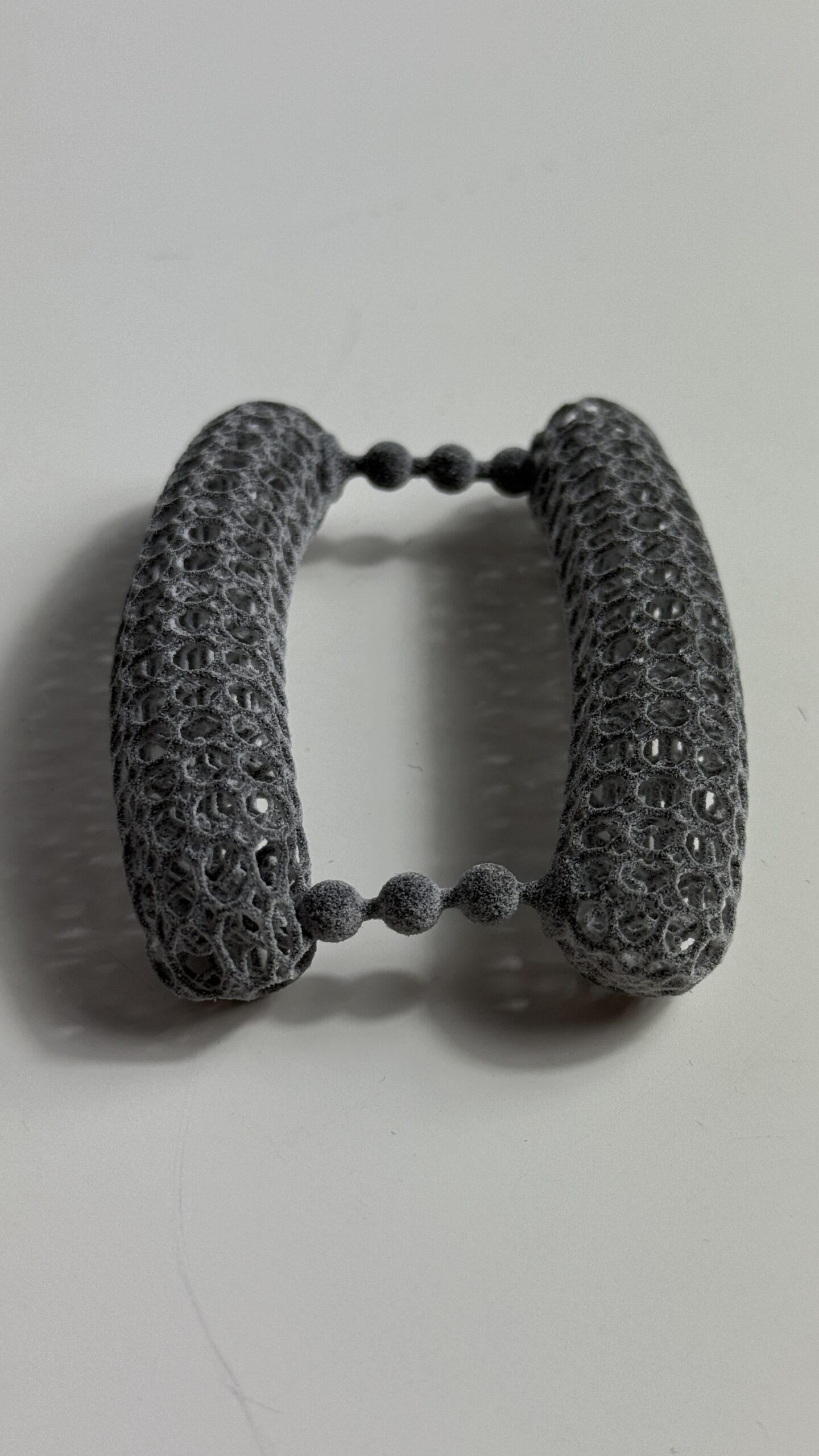
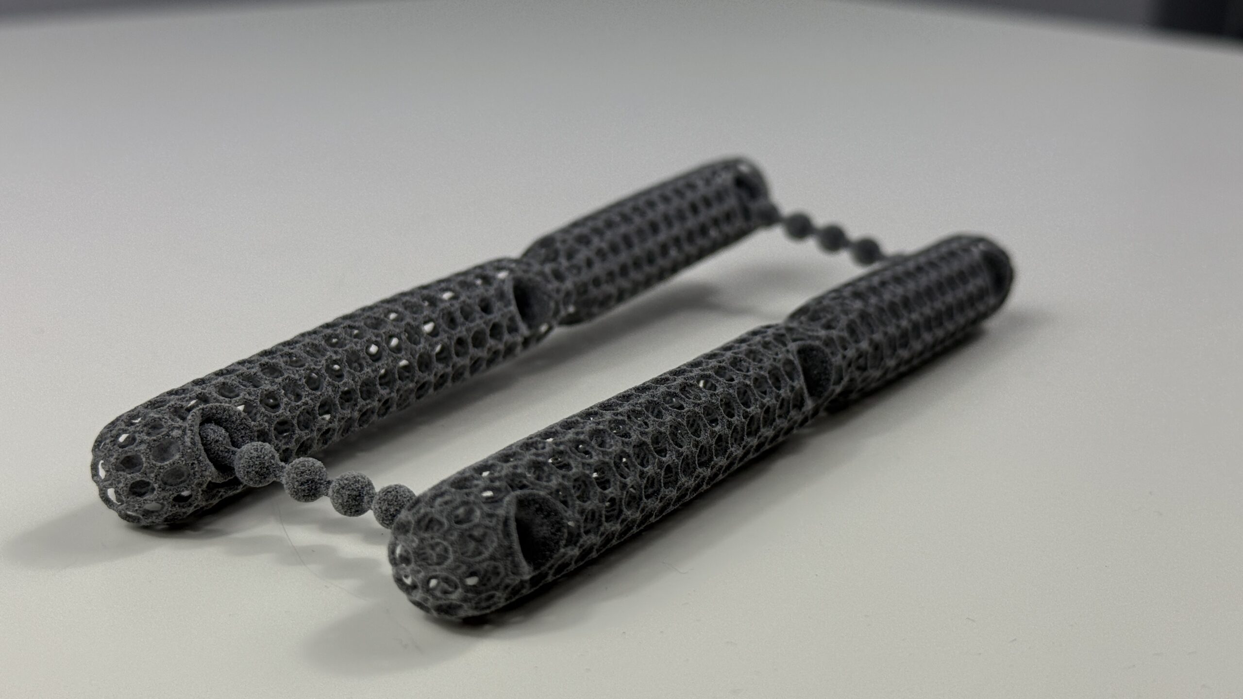
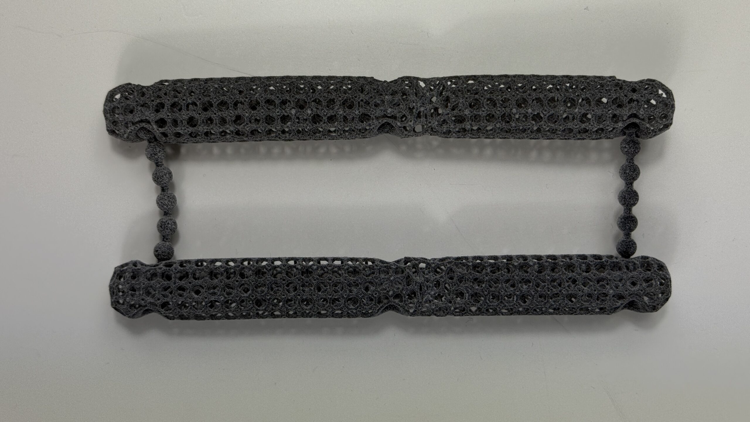
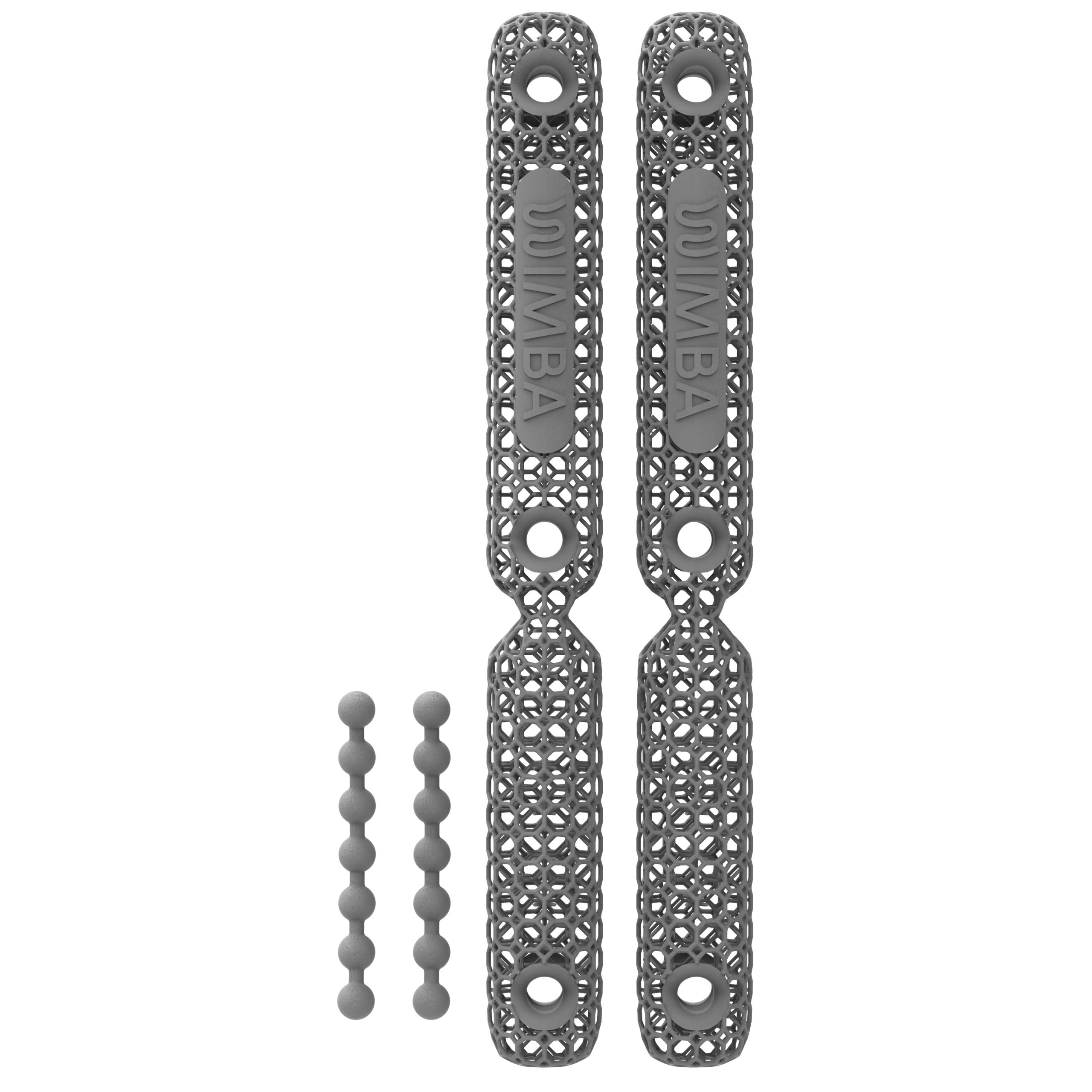
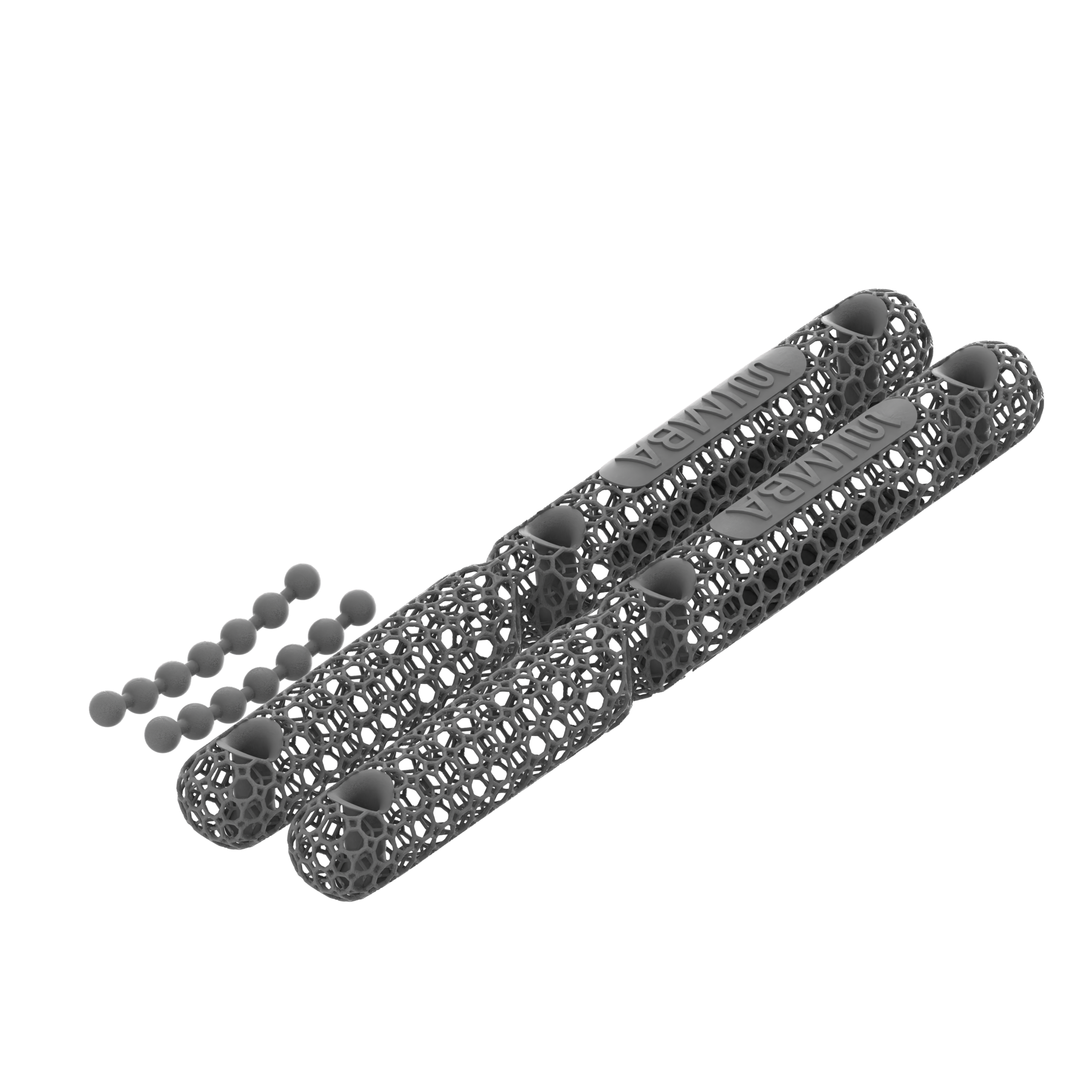
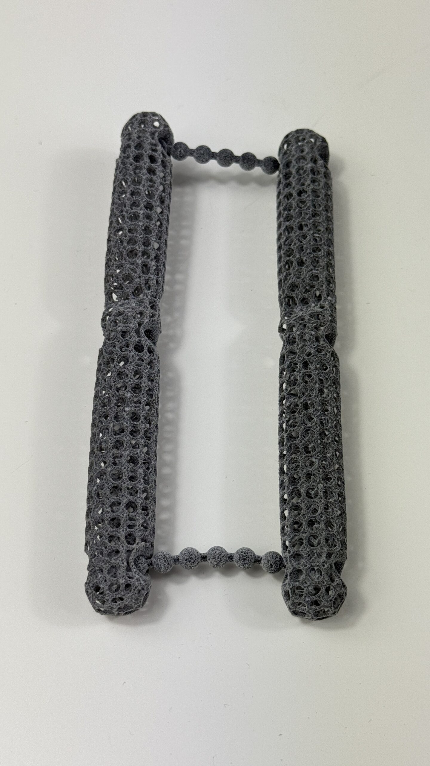
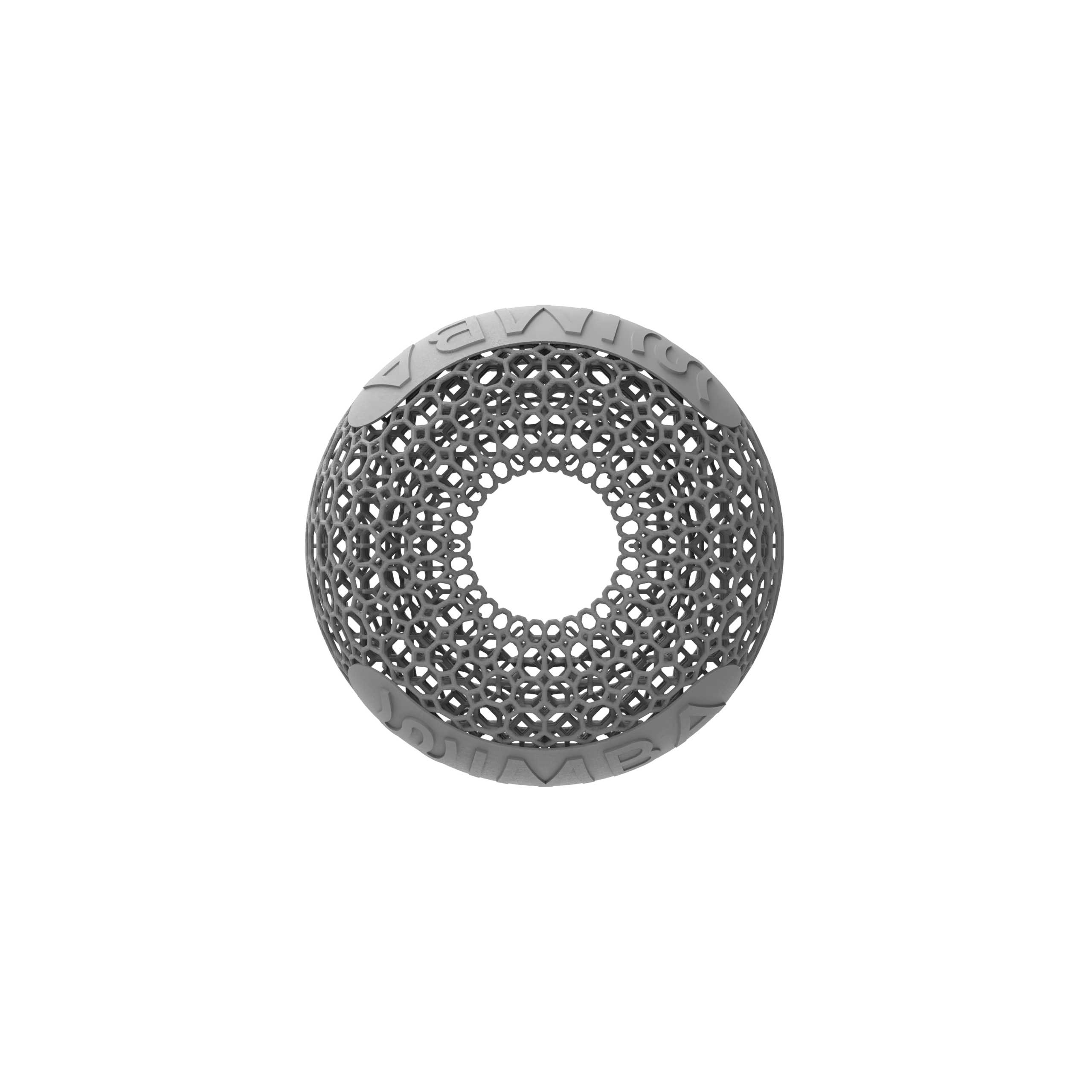
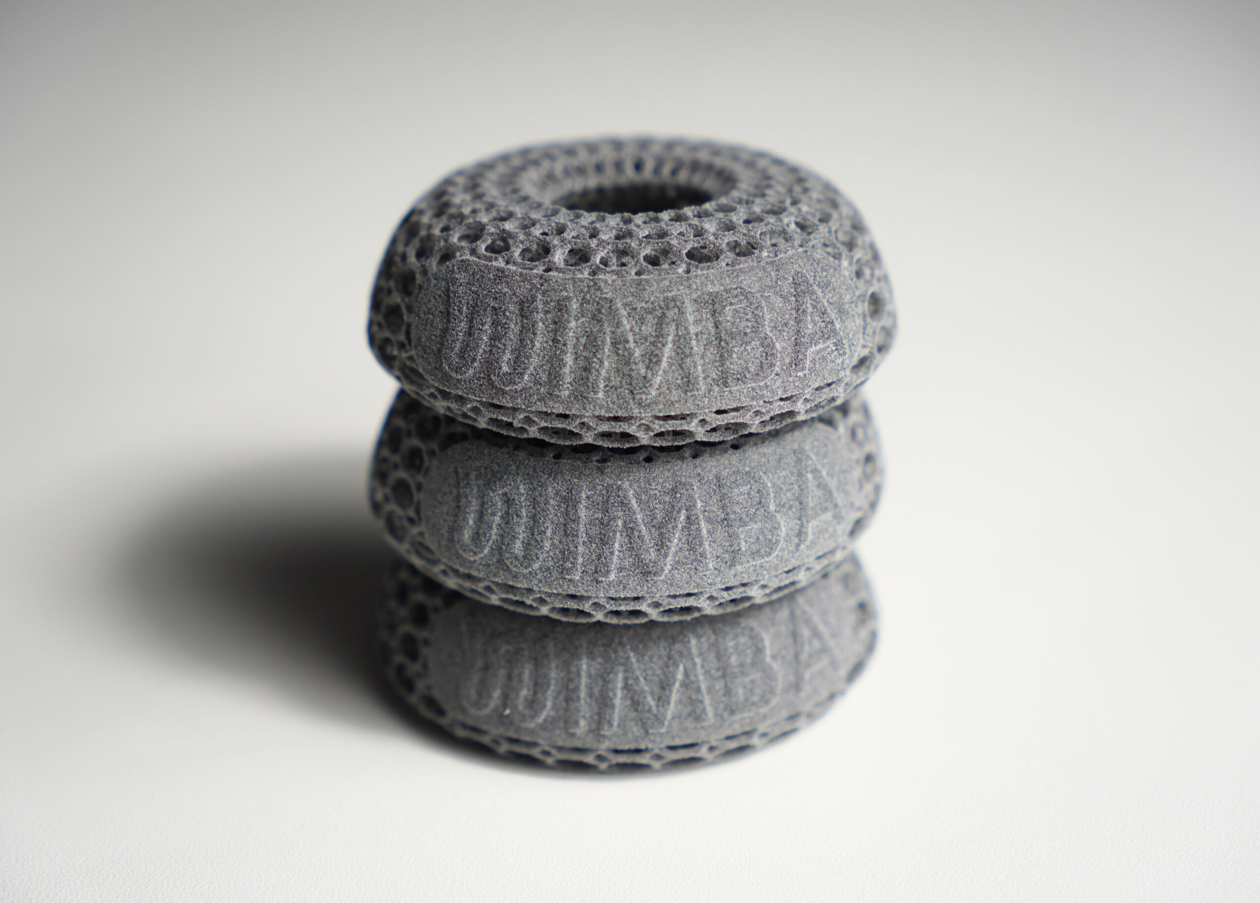
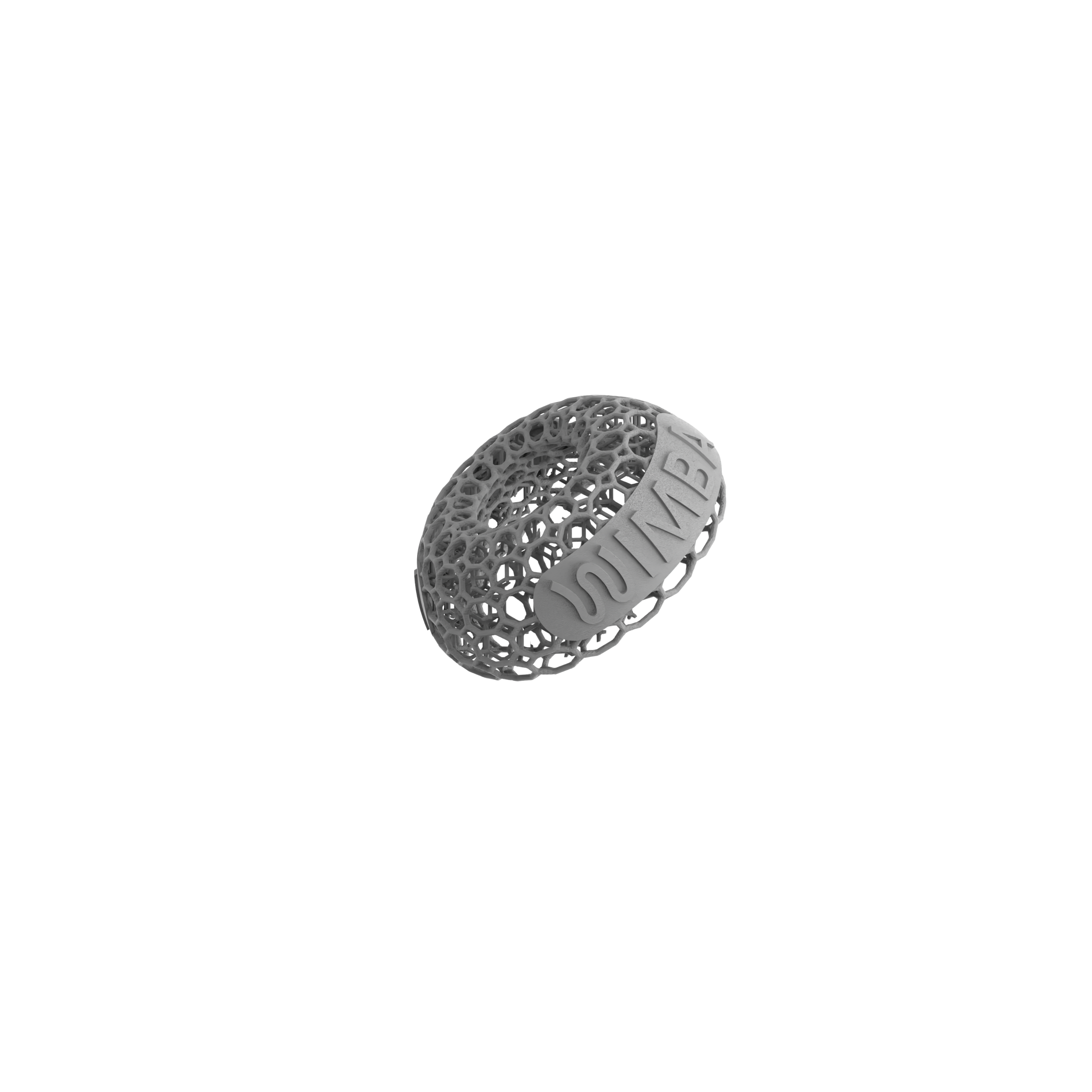
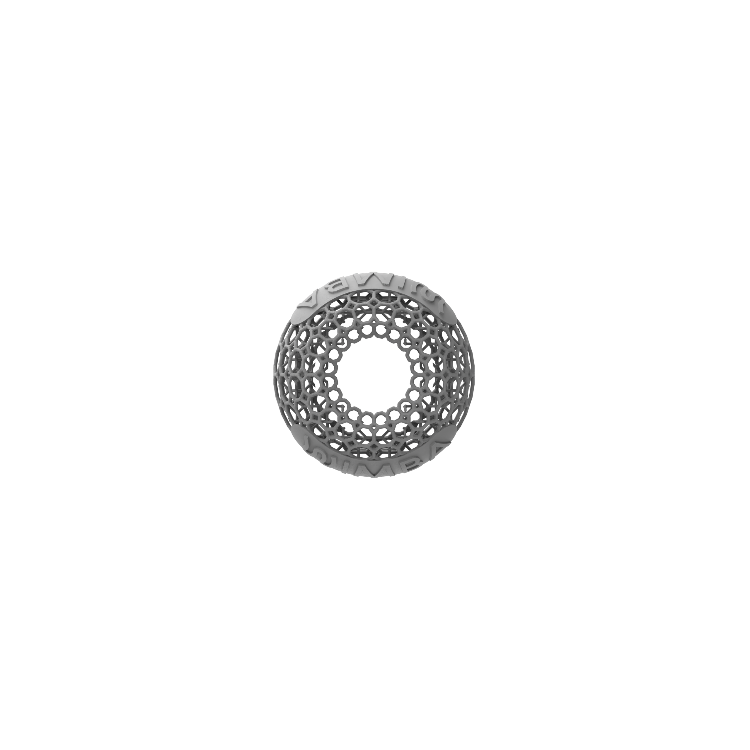
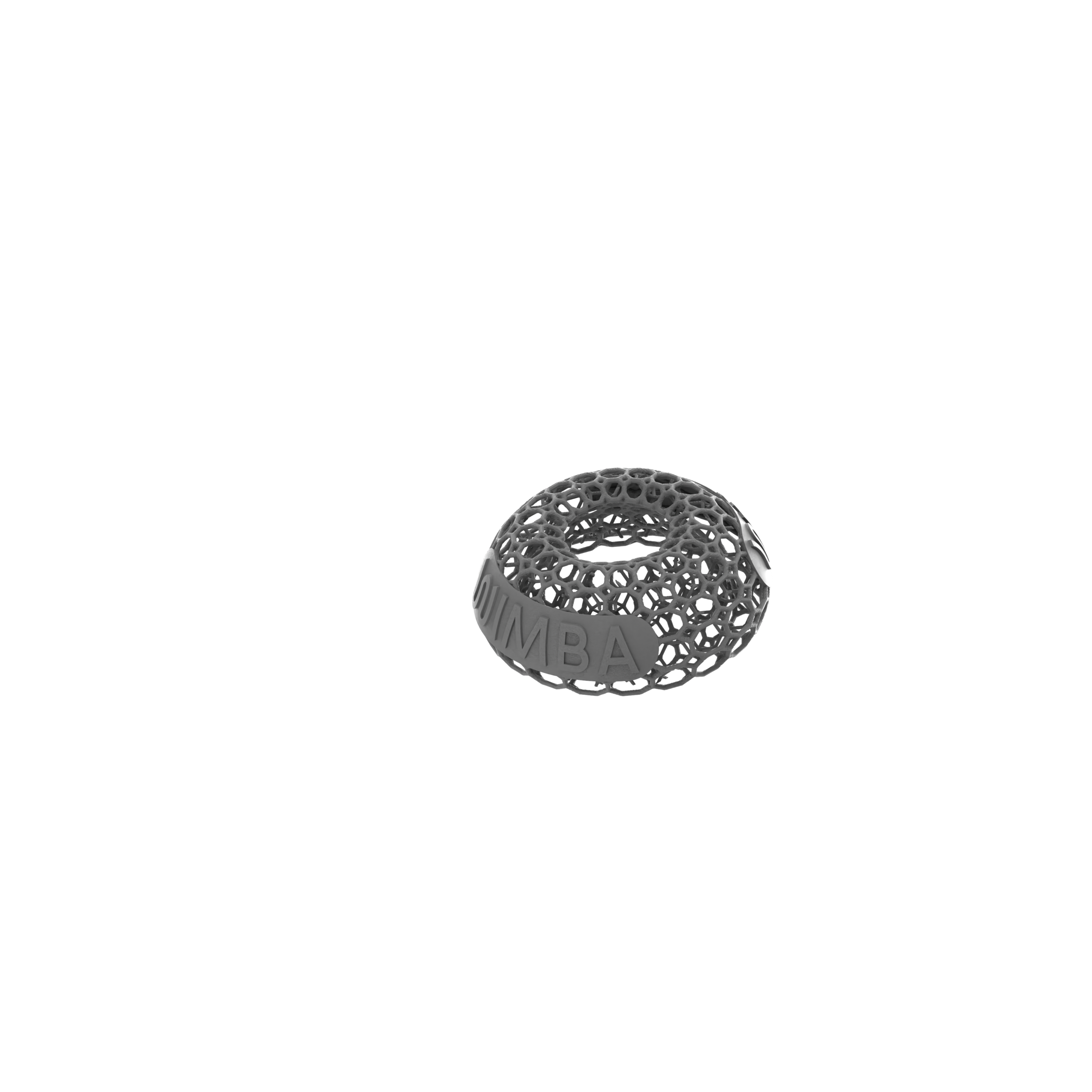
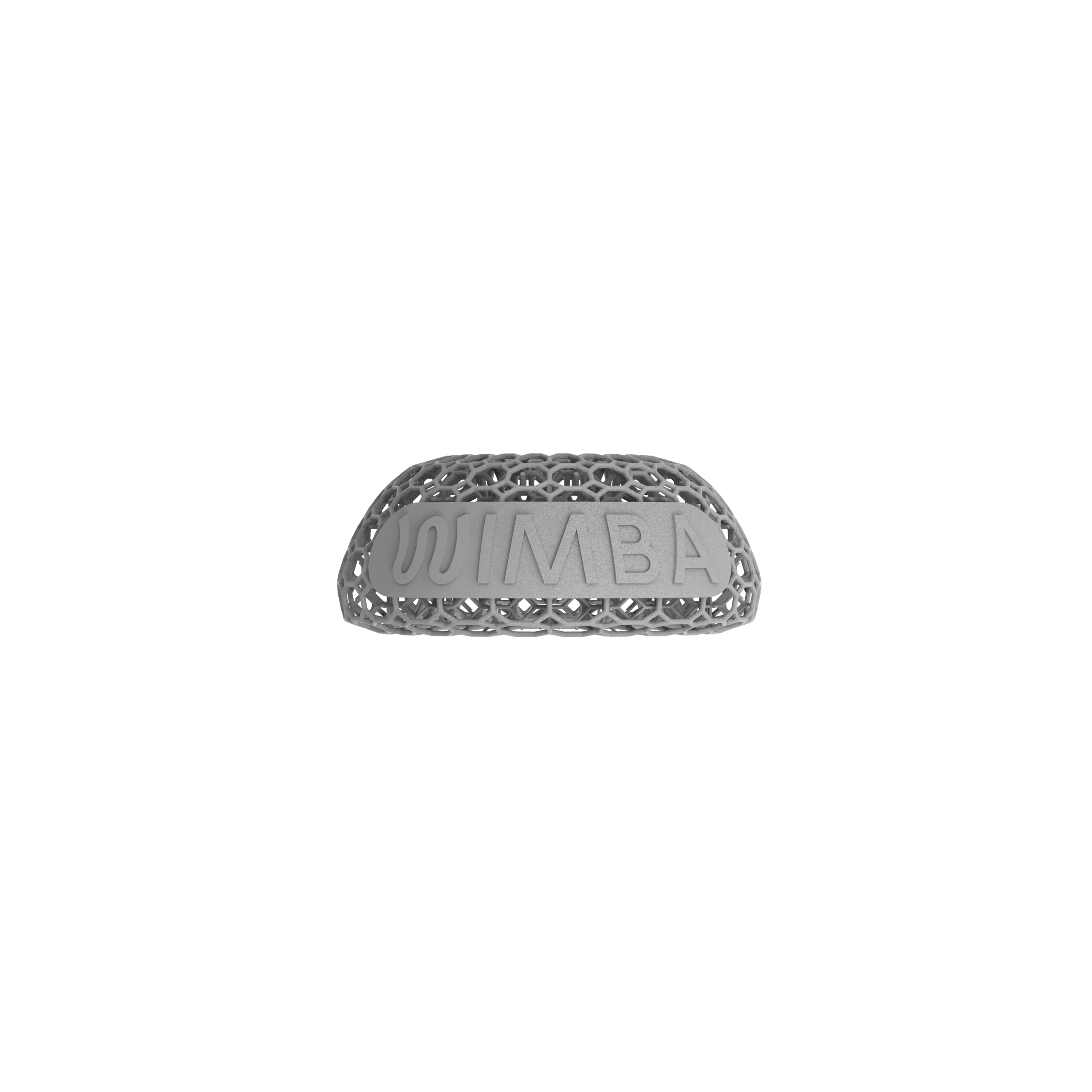
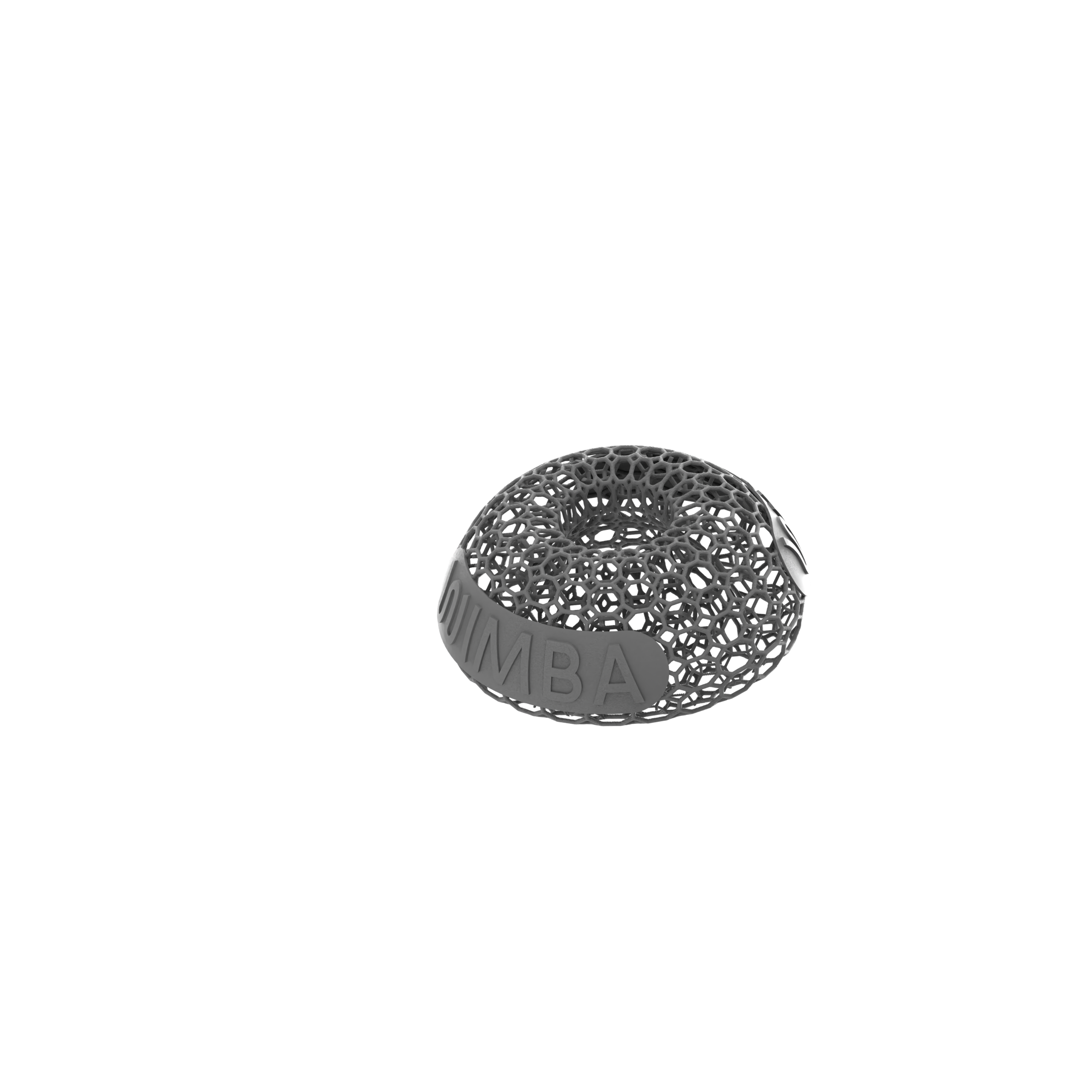
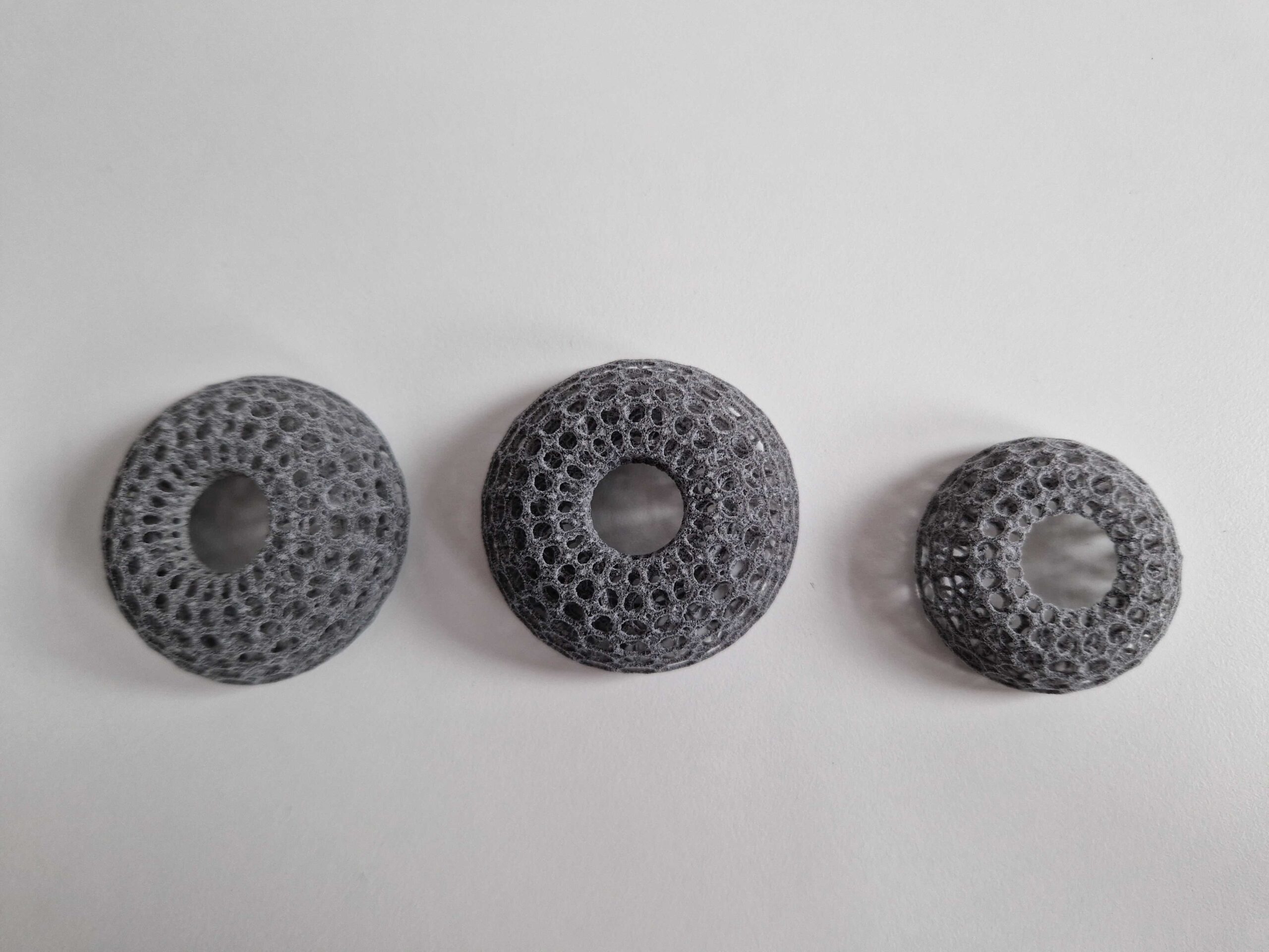
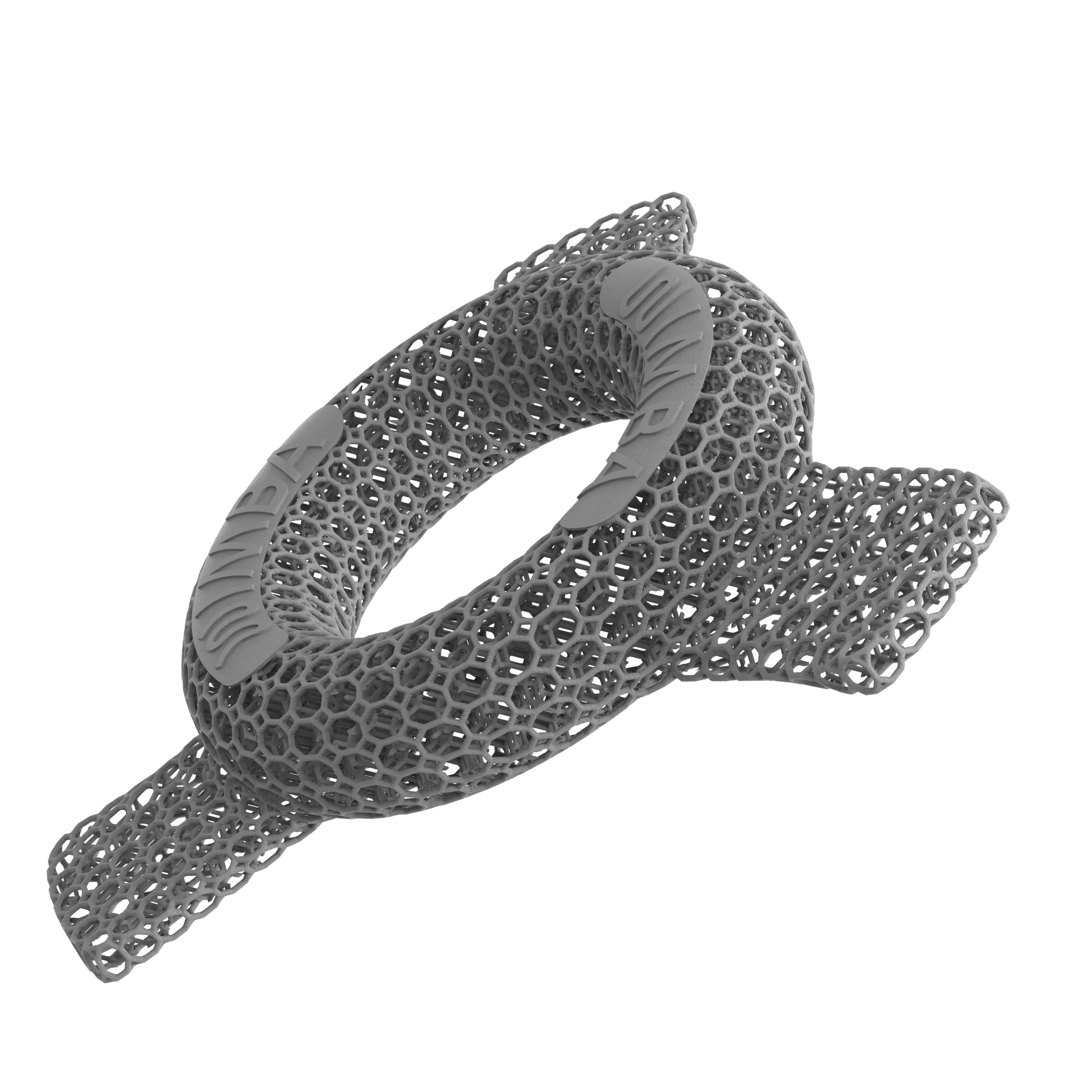
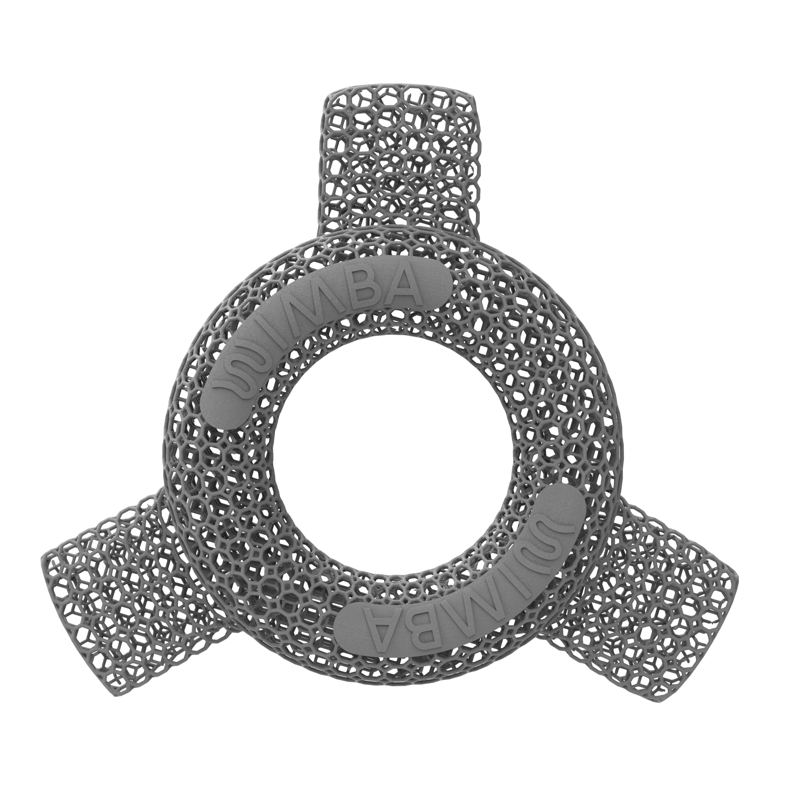
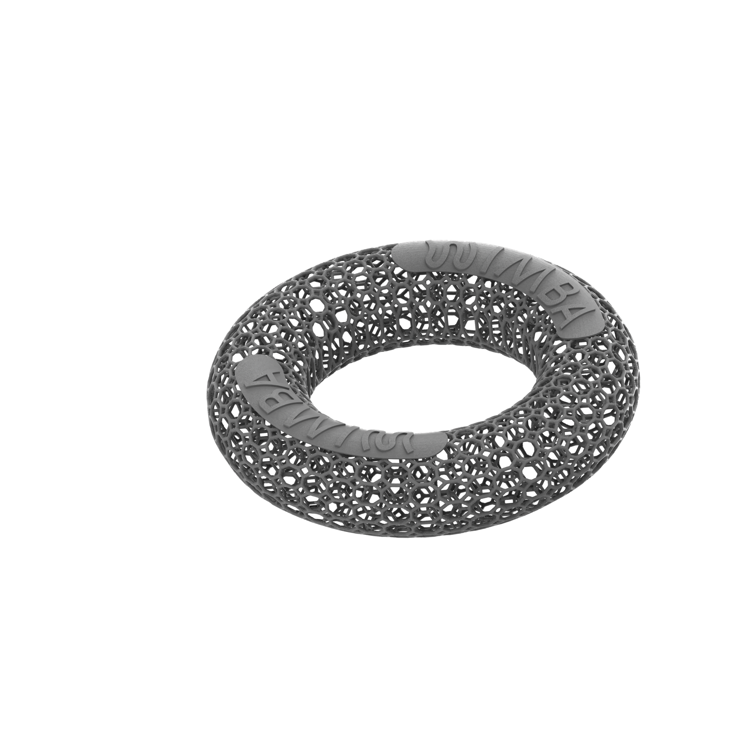
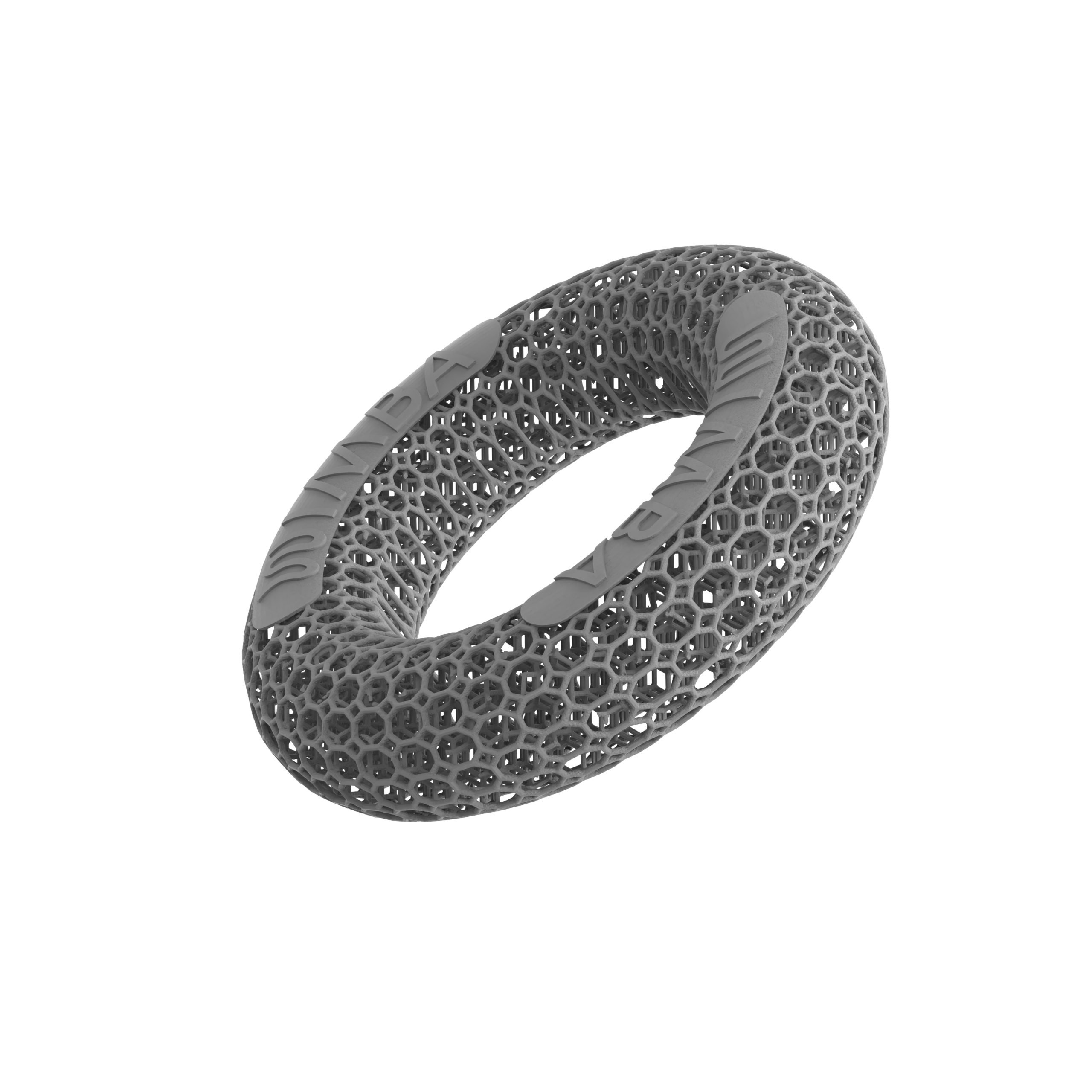
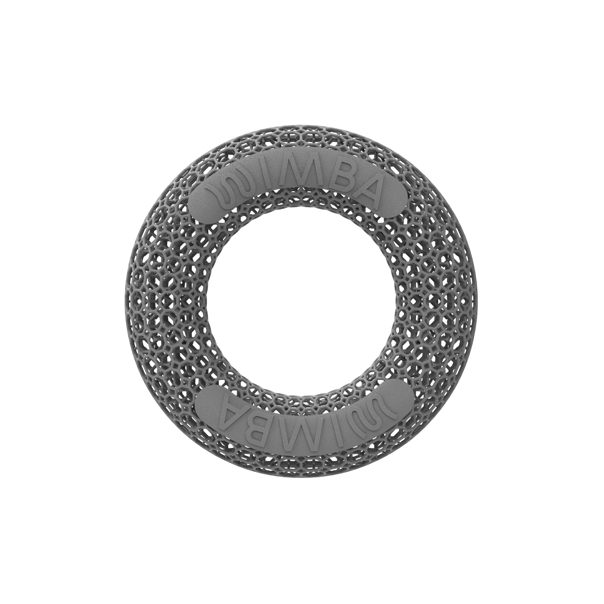
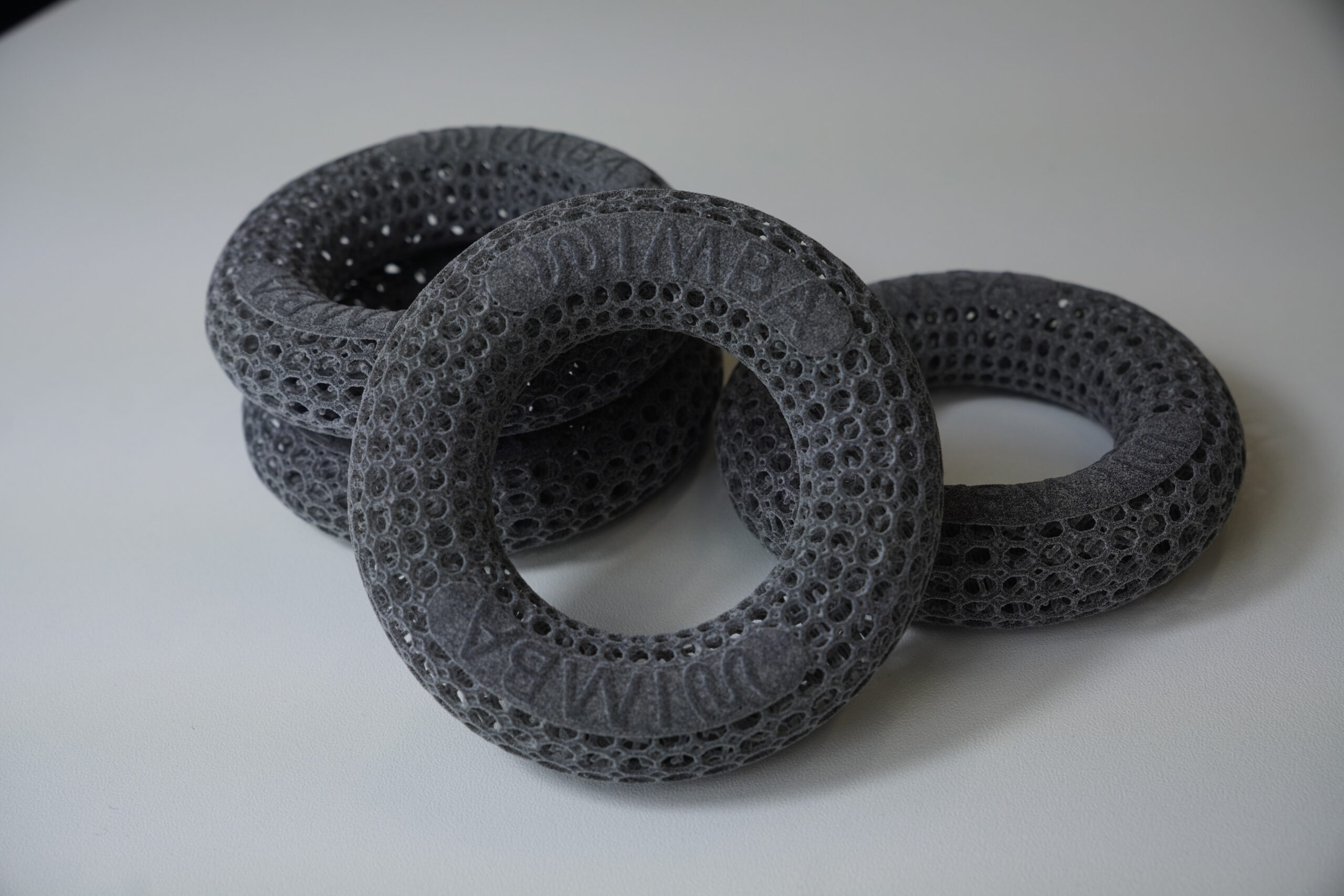
WIMBA custom devices are fabricated from a 3D scan of each patient’s limb using HP Multi Jet Fusion technology. This produces a device accurate to ±0.3 mm, with internal lattice structures that balance rigidity and ventilation. Fit is verified before dispatch and adjusted at no charge if issues arise.
For clinical consultations on device selection for specific patients, register as a WIMBA Provider — case consultation is free for all registered veterinary professionals.