 Skip to content

Understanding and Managing Hip Dysplasia in Dogs
Hip dysplasia in dogs is one of the most common orthopedic conditions veterinary teams diagnose, and one of the most misunderstood by pet owners. A diagnosis can feel overwhelming, especially when it arrives alongside talk of surgery, lifelong medication, or a dog that seems to be slowing down years before its time. The good news is that canine hip dysplasia is well studied, and many affected dogs can maintain a good quality of life with the right combination of veterinary care, weight management, rehabilitation, and, when a separate joint problem is present, carefully selected orthotic support.
This guide to hip dysplasia in dogs explains how the condition develops, how it is diagnosed, which surgical and conservative treatment paths may be considered, and where custom orthotic devices for other joints can — and cannot — fit into a broader management plan.
Could a joint brace be part of your dog’s hip dysplasia plan?
Hip dysplasia often affects how a dog moves overall. Find a WIMBA provider near you for a professional assessment of whether a custom brace could support your dog’s other joints.
⚠️ An important distinction: WIMBA does not manufacture a hip brace. No external orthosis can reshape the acetabulum, restore normal hip congruity, or correct the laxity responsible for hip dysplasia in dogs. A stifle or tarsal orthosis should be considered only when a veterinary examination identifies a separate problem in that joint — not simply because hip dysplasia is present.
What Is Hip Dysplasia in Dogs?
Hip dysplasia is a developmental disorder of the coxofemoral joint. In a normally formed hip, the head of the femur sits securely within the acetabulum, allowing stable movement. In a dysplastic hip, abnormal joint laxity permits excessive movement and subluxation. Repeated instability can contribute to cartilage damage, bone remodeling, inflammation, and osteoarthritis over time. In a four-breed cohort, the PennHIP distraction index — a measure of passive hip laxity — was the most important evaluated risk factor for later hip osteoarthritis.[2]
Hip dysplasia in dogs is complex rather than a single-gene disorder. Genetic research in more than 700 German Shepherd Dogs identified different loci associated with joint incongruity, dysplasia severity, and osteoarthritis, reinforcing that several biological pathways contribute to the phenotype.[1] Growth, body condition, nutrition, and other environmental influences can also affect how the condition is expressed.
How Hip Dysplasia Develops
Hip dysplasia in dogs develops during growth. When the soft tissues surrounding the hip do not keep the femoral head securely seated, repeated loading can allow partial displacement within the joint. Over time, abnormal contact and instability may alter the shape of the acetabulum and femoral head and promote osteoarthritic change.
Hip dysplasia in dogs is reported frequently in many large and giant breeds, but it can affect dogs of any size. Clinical signs do not always match radiographic severity: some young dogs are uncomfortable before substantial arthritis is visible, while other dogs with marked radiographic change show relatively subtle signs. That is why treatment decisions should be based on the individual dog’s pain, function, examination, and imaging rather than an X-ray grade alone.
Common Signs to Watch For
Signs of hip dysplasia in dogs can be subtle at first and may worsen gradually. Owners and veterinary teams commonly look for:
- A “bunny hopping” gait, with both hind limbs moving together
- Reluctance or difficulty rising from a lying or sitting position
- Stiffness after rest that improves somewhat with movement
- Reduced tolerance for exercise, jumping, or climbing stairs
- Loss of muscle mass over the hindquarters and thighs
- A swaying or loose movement through the pelvis
- Intermittent or persistent hind-limb lameness
These signs overlap with stifle disease, spinal disorders, iliopsoas injury, and other orthopedic or neurological conditions. A veterinary assessment is essential before assuming the hips are responsible.
How Hip Dysplasia in Dogs Is Diagnosed
Diagnosing hip dysplasia in dogs begins with medical history, gait observation, and a complete orthopedic examination. The clinician evaluates pain, range of motion, muscle mass, and other joints that could contribute to the dog’s movement pattern. Hip laxity may be assessed under appropriate sedation using techniques such as the Ortolani test.
Imaging and Screening Methods
- Standard hip-extended radiographs assess joint congruity, subluxation, remodeling, and osteoarthritis. Registries such as the Orthopedic Foundation for Animals use this type of view for hip scoring.
- PennHIP radiography uses compression, distraction, and hip-extended views. Its distraction index quantifies passive laxity and can help estimate the risk of later osteoarthritis.[2]
- CT or other advanced imaging may be used in selected cases, especially when more detailed joint assessment or surgical planning is required.
Imaging must be interpreted together with the clinical examination. It also helps distinguish hip dysplasia in dogs from other causes of hind-limb dysfunction and informs which treatment options are realistic for the individual patient.
Treatment Options: Surgical Approaches
Surgical treatment for hip dysplasia in dogs is selected according to age, remaining growth, degree of laxity, existing osteoarthritis, pain, function, and the owner’s goals. These procedures are not interchangeable, and referral to an orthopedic surgeon may be appropriate.
Juvenile Pubic Symphysiodesis
JPS is a preventive growth-modifying procedure for carefully selected, very young puppies. In a small randomized study, surgery at 15 or 20 weeks improved hip conformation and laxity, with greater changes in puppies treated at 15 weeks.[6]
Double or Triple Pelvic Osteotomy
DPO or TPO reorients the acetabulum to improve femoral head coverage. It is generally considered for skeletally immature dogs with laxity but little or no established osteoarthritis.
Femoral Head and Neck Ostectomy
FHO or FHNE removes the femoral head and neck so that a fibrous false joint can form. Outcome depends on factors including body size, muscle condition, rehabilitation, and the individual dog’s function.
Total Hip Replacement
THR replaces the painful joint with prosthetic components. In a prospective force-plate study of 16 dogs with hip dysplasia and secondary degenerative joint disease, loading of the treated limb improved substantially by three to six months after surgery.[7]
Surgery for hip dysplasia in dogs is not appropriate, necessary, or accessible for every patient. Conservative management remains central for non-surgical patients and continues to matter before and after surgery.
Conservative Management: The Foundation of Long-Term Care
For many patients, conservative management is the day-to-day foundation of living with hip dysplasia in dogs. The plan should be individualized and reviewed as pain, function, body condition, and activity change.
Weight Control
Maintaining a lean body condition reduces unnecessary mechanical load, making weight control one of the most practical long-term priorities when managing hip dysplasia in dogs. In a lifelong paired-feeding study of Labrador Retrievers, diet restriction delayed and reduced radiographic hip osteoarthritis.[3] In a separate clinical study, weight reduction improved lameness in overweight dogs with hip osteoarthritis.[4] A veterinarian should set safe calorie and body-condition targets for each patient.
Exercise Modification
Regular, controlled activity helps preserve muscle and mobility without repeated overload. The exact plan may include measured leash walks, gradual strengthening, reduced jumping, and adjustments to duration or surface. Consistency is generally preferable to occasional bursts of strenuous activity followed by prolonged rest.
Physiotherapy and Aquatic Exercise
A rehabilitation professional may use strengthening, balance work, range-of-motion exercises, and aquatic therapy according to the dog’s tolerance. A small kinematic study showed that swimming changes joint motion and produced greater hip range of motion than walking in healthy dogs, but it did not test clinical outcomes in dogs with hip dysplasia.[5] Hydrotherapy should therefore be prescribed for a defined goal rather than treated as a universal solution.
Pain Relief and Medical Management
The veterinarian may recommend prescription analgesia or anti-inflammatory medication and may discuss selected nutritional supplements. Product choice, dosing, monitoring, and laboratory checks should be based on the individual dog’s health and concurrent medication. Owners should not substitute human pain medication for veterinary treatment.
What the Published Evidence Actually Shows
Research supports hip laxity assessment, lean body condition, selected early surgery, and total hip replacement in appropriately chosen patients.[2][3][6][7] These studies do not establish a stifle or tarsal orthosis as a treatment for hip dysplasia in dogs. Distal-limb bracing has a different purpose and requires its own diagnosis and clinical objective.
Where Orthotic Support for Other Joints Fits In
When managing hip dysplasia in dogs, it is important to recognize that an external device cannot stabilize the coxofemoral joint itself because the hip lies deep within the pelvis and surrounding musculature. The presence of hip dysplasia in dogs does not automatically justify bracing another joint. Orthotic support becomes relevant only when the dog’s altered movement is accompanied by a diagnosed problem in a more accessible joint such as the stifle or tarsus.
Dogs with painful hips may redistribute load and change how they use the hind limbs or forelimbs, but compensation alone does not identify which structure needs support. A large retrospective study found that hip dysplasia and cranial cruciate ligament deficiency share several population-level risk factors; it did not prove that hip dysplasia directly causes cruciate disease.[8] Any new lameness therefore needs its own orthopedic workup.
Stifle Orthosis
A custom Stifle Orthosis may be considered when examination confirms a suitable stifle indication, such as selected cases of cranial cruciate ligament insufficiency. It is not prescribed merely to unload a dysplastic hip.
Tarsal Orthosis
A custom Tarsal Orthosis may support an independently diagnosed hock instability, hyperextension, osteoarthritis, or other current product indication. It does not correct hip laxity.
When another joint genuinely requires support, an orthosis can form one part of the plan alongside weight control, pain management, activity modification, and rehabilitation. It should not delay appropriate treatment of the hips or replace surgery when surgery is clinically indicated.
How WIMBA’s Custom Orthotic Process Works
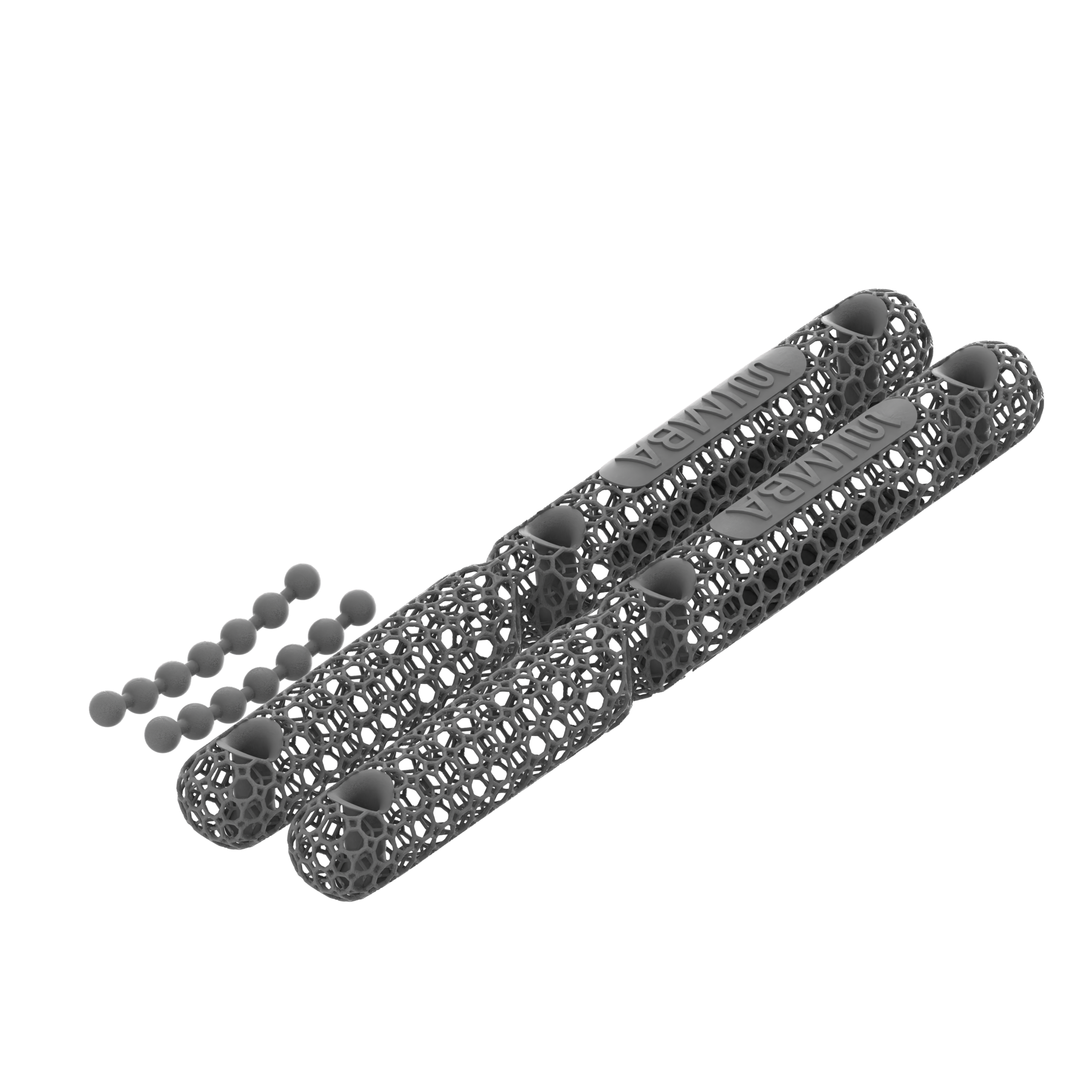
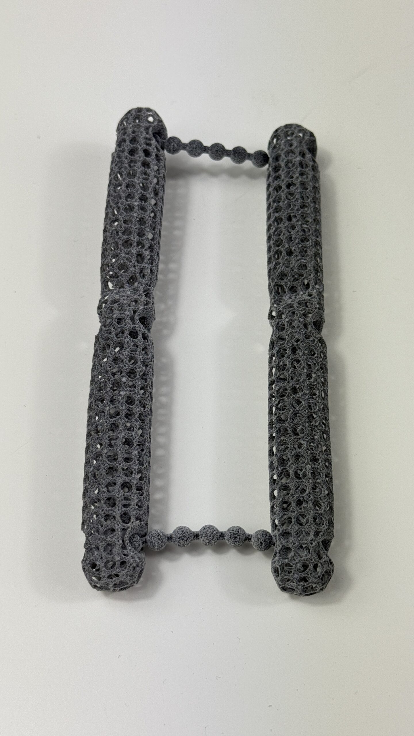
If the veterinary assessment identifies a separate stifle or tarsal indication, WIMBA builds the device around that joint and that patient. The data-capture pathway depends on whether the selected product belongs to the GO or PRO range:
- Confirm the indication and goal. The provider assesses the patient and determines whether a current WIMBA indication matches the diagnosed problem. Uncertain or complex cases can be submitted for a WIMBA Case Consult.
- Choose the correct data pathway. Eligible GO products use product-specific photographs and manual measurements submitted through WimbaAPP. PRO devices, including WIMBA Stifle Orthosis Pro, require manual measurements and a WimbaSCAN performed through the applicable Provider Pro workflow.
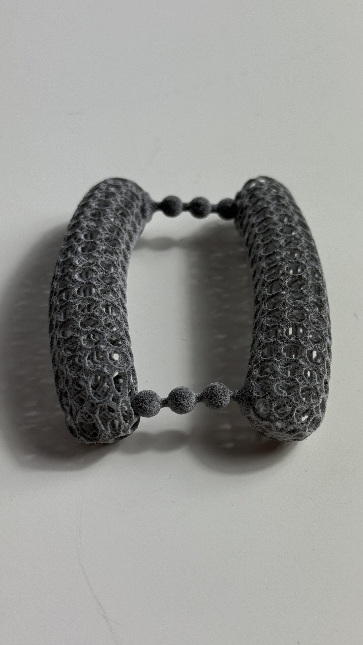
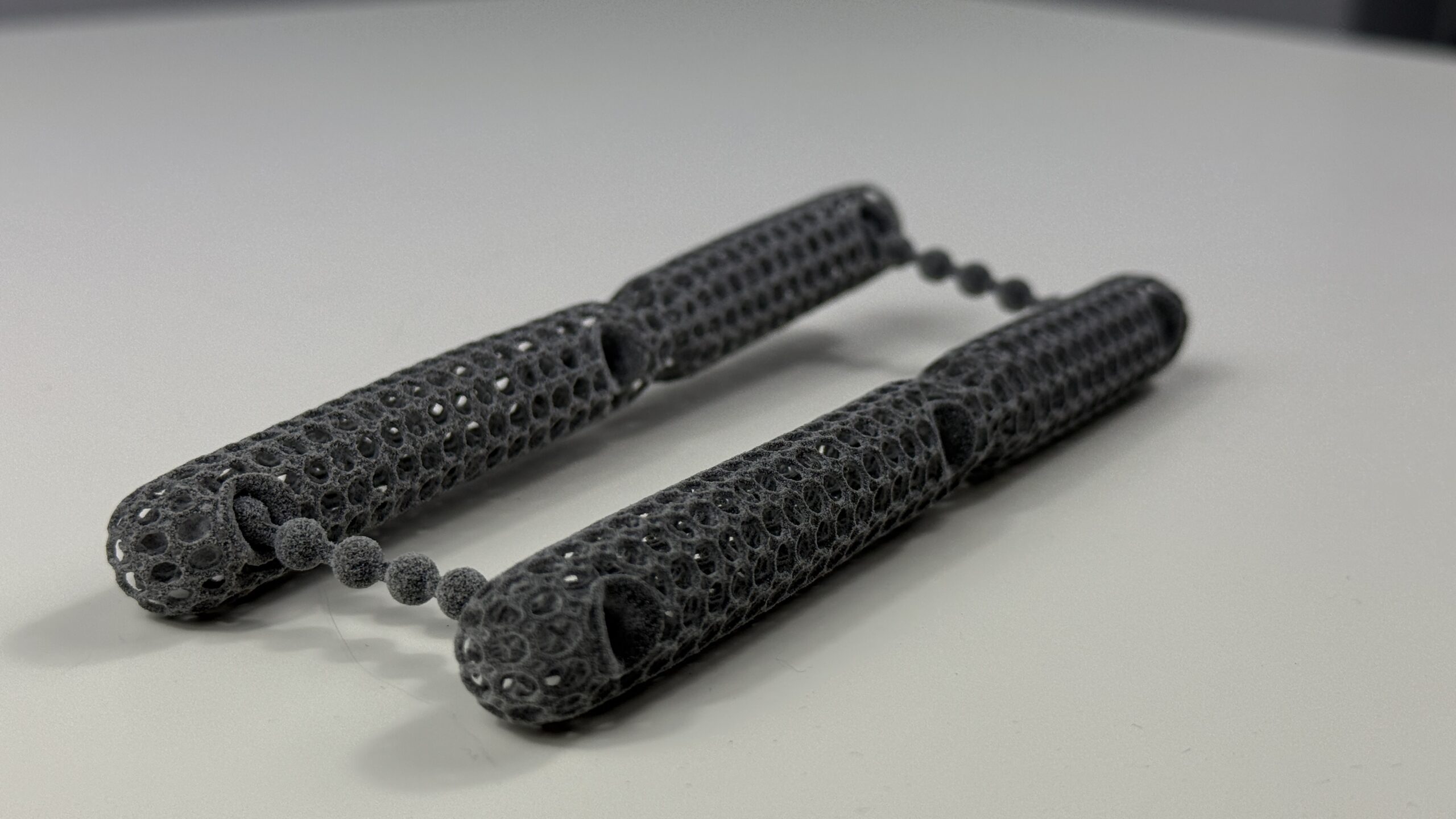
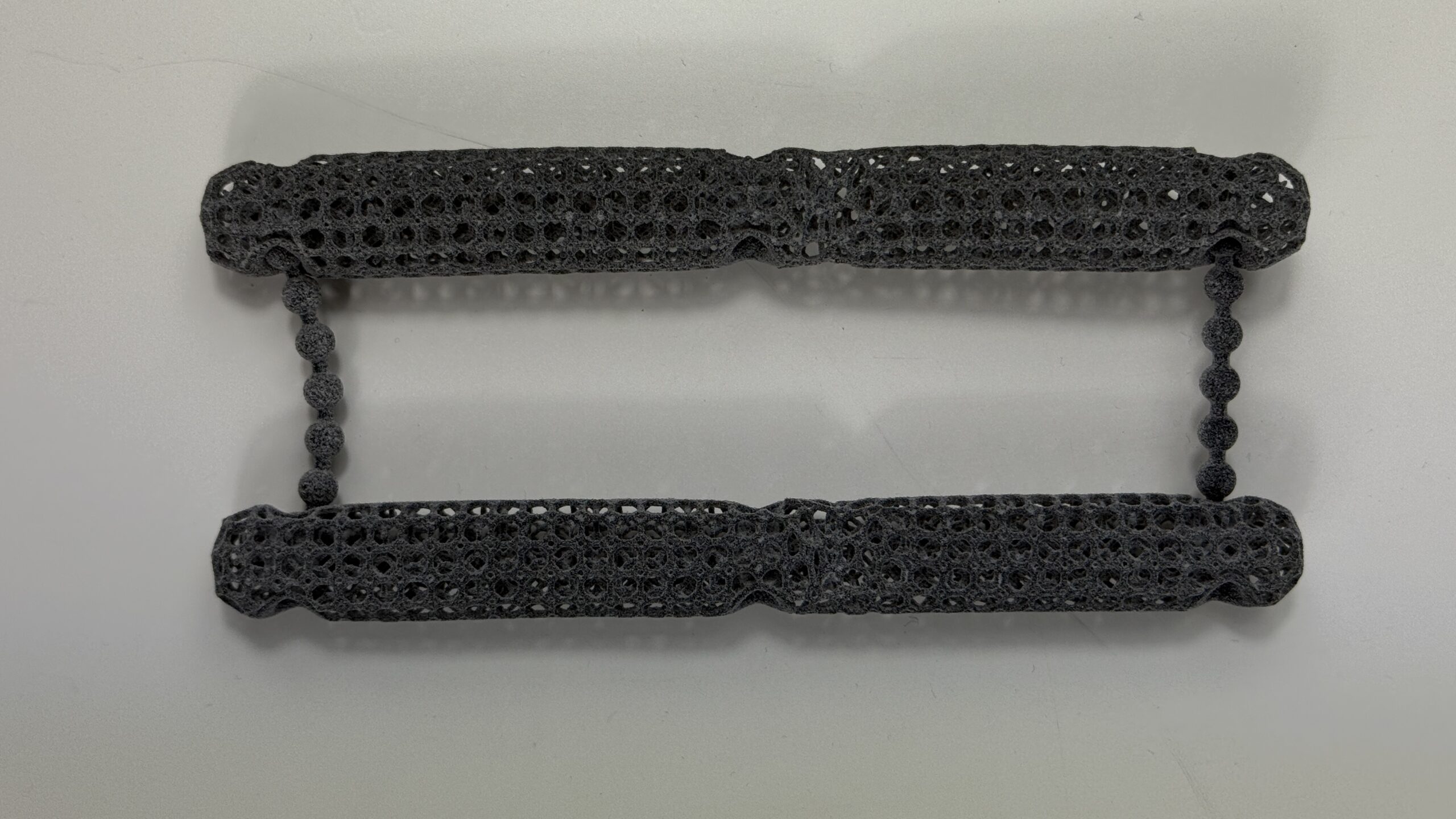
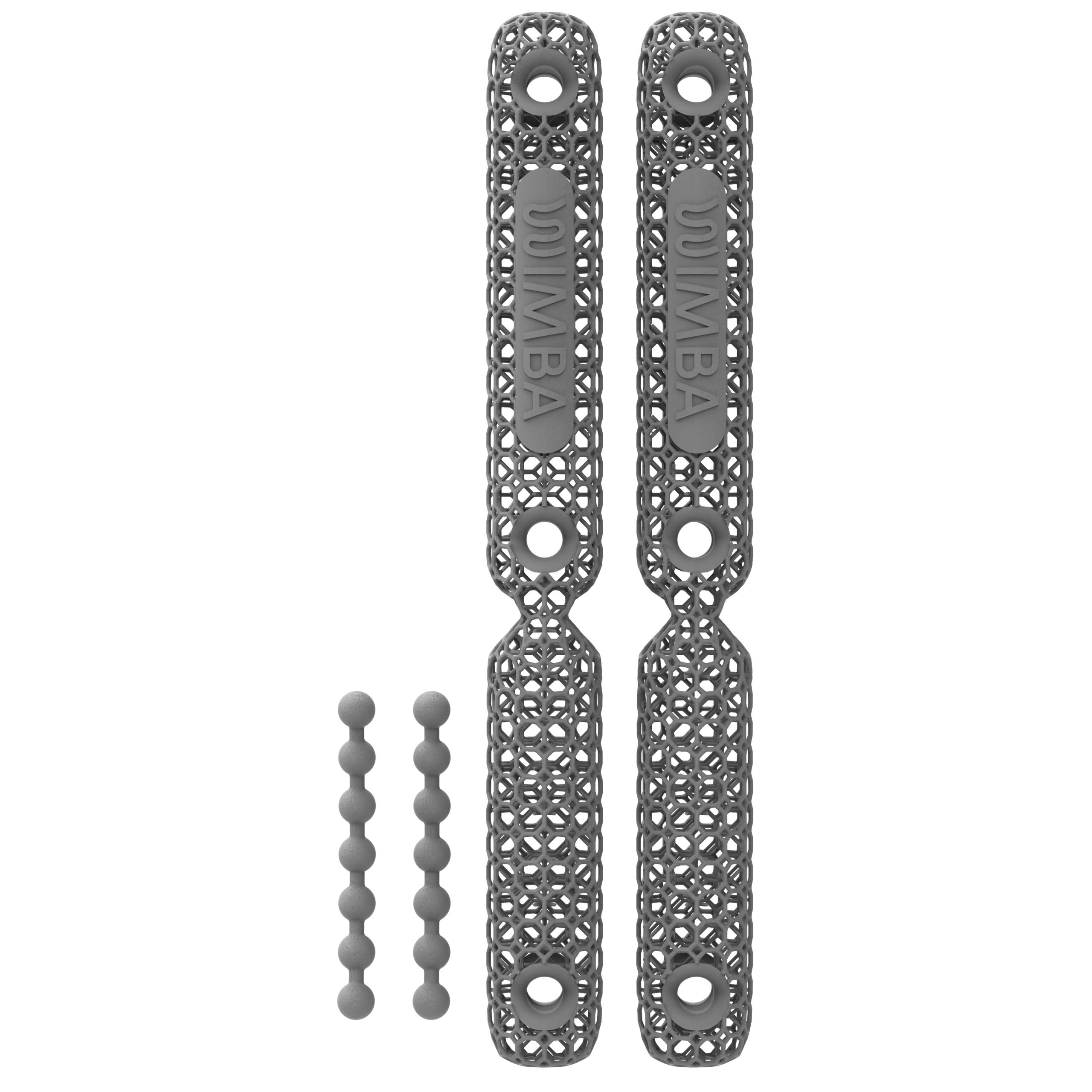
- Design and manufacture the device. Patient-specific data are used to create the digital design. The orthosis is manufactured using HP Multi Jet Fusion 3D printing for a lightweight, durable structure.
- Fit and follow up through the veterinary team. The provider checks alignment, suspension, skin contact, comfort, and controlled movement, then teaches the owner how to apply, remove, clean, and monitor the device.
⚠️ Fit and monitoring matter: Prospective canine data show that skin problems, mechanical issues, and patient non-acceptance can occur with veterinary orthoses.[9] Introduce the device according to the provider’s schedule, check the skin before and after wear, and request reassessment if rubbing, redness, device movement, distress, pain, or worsening gait appears.
Frequently Asked Questions
Can an orthosis cure or reverse hip dysplasia in dogs?
No. Hip dysplasia is a structural condition of the hip, and an external orthosis cannot correct hip conformation or joint laxity. WIMBA devices may support a separately diagnosed problem in the stifle or tarsus; they do not treat the dysplastic hip itself.
Does WIMBA make a hip brace?
No. WIMBA does not currently manufacture a hip brace. Its orthotic range supports joints such as the stifle, tarsus, and carpus when the veterinary assessment confirms a suitable indication.
Is surgery always necessary for hip dysplasia?
No. Some dogs are managed successfully with weight control, appropriate exercise, rehabilitation, and veterinary pain management. Surgery may be recommended when age, anatomy, pain, functional limitation, or disease progression makes it the most appropriate option.
How do I know whether another joint needs an orthosis?
A veterinarian or rehabilitation professional must examine the dog and diagnose the specific joint problem. A changed gait or new lameness does not by itself show whether the stifle, tarsus, hip, spine, or another structure is responsible.
At what age can hip dysplasia in dogs be diagnosed?
Hip laxity can be assessed in young puppies with appropriate examination and imaging, while some dogs are not diagnosed until osteoarthritis becomes symptomatic later in life. Early assessment matters because growth-modifying procedures such as juvenile pubic symphysiodesis are available only during a narrow age window.
Will my dog need to wear an orthosis for life?
That depends on the separate joint condition for which the device was prescribed. Some dogs use an orthosis during a defined rehabilitation period, while others need longer-term support. The veterinary team should reassess fit, skin health, function, and the ongoing clinical goal.
Can WIMBA orthotics manage hip dysplasia day to day?
Not directly. WIMBA does not manufacture a hip brace, and its carpal, tarsal, and stifle orthoses do not stabilize or correct a dysplastic hip. A WIMBA orthosis may be considered only when a veterinary assessment identifies a separate problem in one of those supported joints. Day-to-day hip dysplasia management should follow an individualized plan that may include weight control, appropriate exercise, rehabilitation, veterinary pain management, and surgery when indicated.
What non-surgical options exist for dogs with hip dysplasia?
Non-surgical management is individualized and may include maintaining a lean body condition, veterinarian-directed exercise modification, rehabilitation, and prescribed pain or osteoarthritis treatment. In a small prospective study, weight reduction was associated with reduced lameness in overweight dogs with hip osteoarthritis, but there is no single protocol for every dog. WIMBA does not make a hip brace, and its devices do not treat the dysplastic hip. A WIMBA orthosis may be considered only if a veterinary assessment identifies a separate problem in the carpus, tarsus, or stifle and defines a specific support goal.
How does physical therapy help dogs with hip dysplasia?
A rehabilitation professional may use structured exercises — strengthening, balance work, controlled range-of-motion work, and sometimes aquatic therapy — tailored to the dog’s tolerance and stage of disease. Physical therapy does not correct hip laxity or joint conformation, but it can help preserve muscle mass and support day-to-day function alongside weight control and veterinary pain management. As with other conservative measures, it works best as part of an individualized plan built with a veterinarian or rehabilitation specialist, not as a stand-alone fix.
Getting Started
Hip dysplasia in dogs is a lifelong condition to manage rather than a single problem with one universal solution. The right plan depends on age, pain, function, imaging, other joints involved, concurrent disease, and the owner’s goals. If your dog has been diagnosed with hip dysplasia and develops new strain or lameness elsewhere, ask your veterinarian or rehabilitation specialist to assess the entire movement chain before considering an orthosis.
Explore real device applications in Patient Stories. Veterinary professionals can submit a patient case for guidance, while pet owners can use the directory to find a WIMBA Provider.
This article is for general educational purposes and is not a substitute for veterinary diagnosis or treatment planning. WIMBA orthotic devices are prescribed and fitted by qualified veterinary professionals as part of an individualized care plan. Always consult your veterinarian regarding hip dysplasia in dogs or any other orthopedic condition in your pet.
Scientific References
- Mikkola L, Holopainen S, Pessa-Morikawa T, et al. Genetic dissection of canine hip dysplasia phenotypes and osteoarthritis reveals three novel loci. BMC Genomics. 2019;20:1027. doi:10.1186/s12864-019-6422-6
- Runge JJ, Kelly SP, Gregor TP, Kotwal S, Smith GK. Distraction index as a risk factor for osteoarthritis associated with hip dysplasia in four large dog breeds. J Small Anim Pract. 2010;51(5):264–269. doi:10.1111/j.1748-5827.2010.00937.x
- Smith GK, Paster ER, Powers MY, et al. Lifelong diet restriction and radiographic evidence of osteoarthritis of the hip joint in dogs. J Am Vet Med Assoc. 2006;229(5):690–693. doi:10.2460/javma.229.5.690
- Impellizeri JA, Tetrick MA, Muir P. Effect of weight reduction on clinical signs of lameness in dogs with hip osteoarthritis. J Am Vet Med Assoc. 2000;216(7):1089–1091. doi:10.2460/javma.2000.216.1089
- Marsolais GS, McLean S, Derrick T, Conzemius MG. Kinematic analysis of the hind limb during swimming and walking in healthy dogs and dogs with surgically corrected cranial cruciate ligament rupture. J Am Vet Med Assoc. 2003;222(6):739–743. doi:10.2460/javma.2003.222.739
- Patricelli AJ, Dueland RT, Adams WM, Fialkowski JP, Linn KA, Nordheim EV. Juvenile pubic symphysiodesis in dysplastic puppies at 15 and 20 weeks of age. Vet Surg. 2002;31(5):435–444. doi:10.1053/jvet.2002.34766
- Budsberg SC, Chambers JN, Lue SL, Foutz TL, Reece L. Prospective evaluation of ground reaction forces in dogs undergoing unilateral total hip replacement. Am J Vet Res. 1996;57(12):1781–1785. PMID:8950435
- Witsberger TH, Villamil JA, Schultz LG, Hahn AW, Cook JL. Prevalence of and risk factors for hip dysplasia and cranial cruciate ligament deficiency in dogs. J Am Vet Med Assoc. 2008;232(12):1818–1824. doi:10.2460/javma.232.12.1818
- Rosen S, Duerr FM, Elam LH. Prospective evaluation of complications associated with orthosis and prosthesis use in canine patients. Front Vet Sci. 2022;9:892662. doi:10.3389/fvets.2022.892662