 Skip to content

Cervical Instability in Dogs: Causes, Signs, Diagnosis, and Support
Cervical instability in dogs means that one or more vertebrae in the neck are not maintaining normal alignment during movement. Depending on the cause and severity, the result may range from neck pain and a guarded posture to weakness, ataxia, or loss of voluntary movement. Because the cervical spinal cord carries signals to all four limbs and the muscles involved in breathing, any suspected instability requires careful veterinary assessment.
Cervical instability in dogs is not one diagnosis and it is not managed with one universal treatment. Congenital malformation, traumatic injury, fracture, and selected upper-cervical disorders can create very different clinical pictures. This guide explains the major causes and warning signs, how veterinarians investigate canine cervical spine instability, and where an external support such as a WIMBA Cervical Collar GO may fit within a veterinarian-directed plan.
Sudden neurological signs are an emergency
Sudden neck pain, wobbliness, weakness, collapse, or inability to stand can indicate spinal cord injury. Keep the dog as still as safely possible and contact a veterinarian or emergency service immediately. Do not flex, extend, test, or brace the neck at home. A dog neck brace should never be applied before the injury has been assessed and the spine has been stabilized by a veterinary professional.
What Causes Cervical Instability in Dogs?
Cervical instability in dogs describes a mechanical problem rather than a single disease. The unstable level, underlying anatomy, degree of spinal cord compression, and speed of onset all influence the signs and treatment options. The following categories should not be treated as interchangeable.
Congenital atlantoaxial instability
The atlantoaxial joint connects the first cervical vertebra, the atlas, to the second, the axis. Malformation or absence of the dens, abnormal ligamentous support, or a combination of abnormalities can permit excessive motion at C1–C2. When cervical instability in dogs arises at this joint, it is called atlantoaxial instability. Atlantoaxial instability in dogs is most often recognized in young miniature and toy breeds, although presentation and anatomy vary. Clinical examination and imaging findings must be interpreted together because measurements alone do not define every clinically relevant case.1
Traumatic upper-cervical injury
Falls, vehicle impacts, bites, and other high-energy events can fracture or displace cervical structures in dogs of any age or size. Traumatic atlantoaxial subluxation may cause anything from severe pain to tetraparesis or tetraplegia. CT can define bony injury and alignment, while MRI may reveal spinal cord and soft-tissue injury that affects clinical decisions.2 Traumatic cervical instability in dogs must be treated as urgent until the extent of injury is known.
Temporary support around surgery
“Post-surgical instability” is not a single disease category. In selected cases, a surgeon may prescribe external support before a procedure, during a controlled recovery period, or after fixation while tissues heal. For cervical instability in dogs during recovery, the device objective, wear schedule, and follow-up plan depend on the procedure and current neurological status. External support must not obscure deterioration or replace the imaging and reassessment required after cervical surgery.
Complex or coexisting conditions
A dog can have hydrocephalus or another neurological disorder at the same time as upper-cervical instability, but coexistence does not prove that one condition caused the other. This distinction matters when discussing cervical instability in dogs. Hydrocephalus by itself is not a standard indication for a cervical collar. Pipi’s WIMBA story involves both hydrocephalus and documented complex cervical instability; it should be understood as one individual case, not a general treatment rule.
Signs of Cervical Instability in Dogs
The clinical picture can be subtle, intermittent, or dramatic. Signs of cervical instability in dogs may reflect pain, spinal cord dysfunction, or both. Owners and veterinary teams may notice:
- neck pain, a guarded posture, or reluctance to turn, raise, or lower the head;
- yelping when the head or neck is touched or moved;
- difficulty reaching food or water, or reluctance to chew because movement is uncomfortable;
- a wobbly, uncoordinated gait affecting two or all four limbs;
- weakness, knuckling, partial paralysis, or loss of voluntary movement;
- sudden collapse or inability to rise after a fall, impact, or rough play;
- changes in breathing, alertness, or neurological function that require immediate emergency assessment.
These signs are not specific to cervical instability in dogs. Intervertebral disc disease, inflammation, infection, neoplasia, and other neurological conditions can produce overlapping signs. That is why a visible symptom should lead to diagnosis rather than an attempt to select a device at home.
How Cervical Instability in Dogs Is Diagnosed
For cervical instability in dogs, diagnosis starts with history, a physical examination, and a neurological examination performed without unsafe manipulation of the neck. The veterinarian localizes the suspected lesion, assesses pain and neurological severity, and determines how the dog can be transported and positioned safely.
Radiographs may help identify gross malformation or displacement, but positioning must be controlled by the veterinary team. CT is particularly useful for bone, fracture configuration, and three-dimensional alignment. MRI adds information about the spinal cord, compression, soft tissues, and concurrent abnormalities. Research in small-breed dogs supports interpreting CT or MRI measurements together with clinical findings rather than treating a single imaging cutoff as a diagnosis.1 A recent surgical cohort also found that MRI abnormalities can contribute prognostic information in atlantoaxial instability in dogs.3
Do not test neck range of motion at home. Flexing or extending an unstable upper-cervical joint can worsen displacement or spinal cord injury. Diagnostic positioning belongs in a controlled clinical setting after the veterinarian has assessed risk.
How External Cervical Support May Help
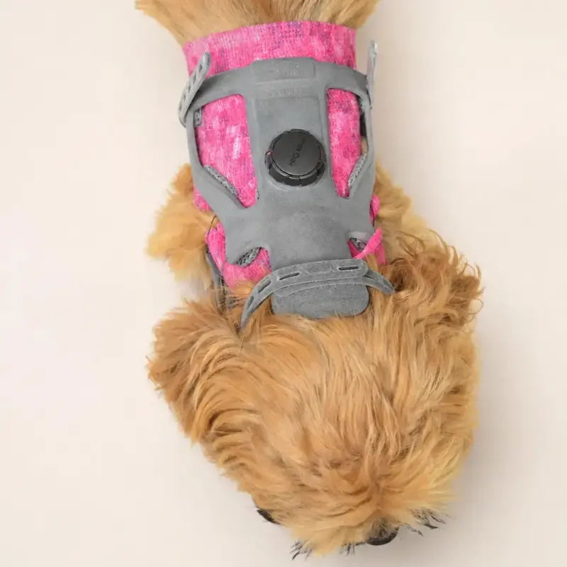
Management of cervical instability in dogs may include medical care, activity restriction, rehabilitation, surgery, external support, or a combination selected for the individual patient. A cervical orthosis for dogs is an external support intended to reduce unwanted movement and help maintain a clinically selected neck position. It does not repair malformed bone, heal a ligament, decompress the spinal cord, or create internal fixation. In carefully selected cases, a dog neck brace may be one component of conservative management, temporary protection around surgery, or comfort-focused support during controlled activity.
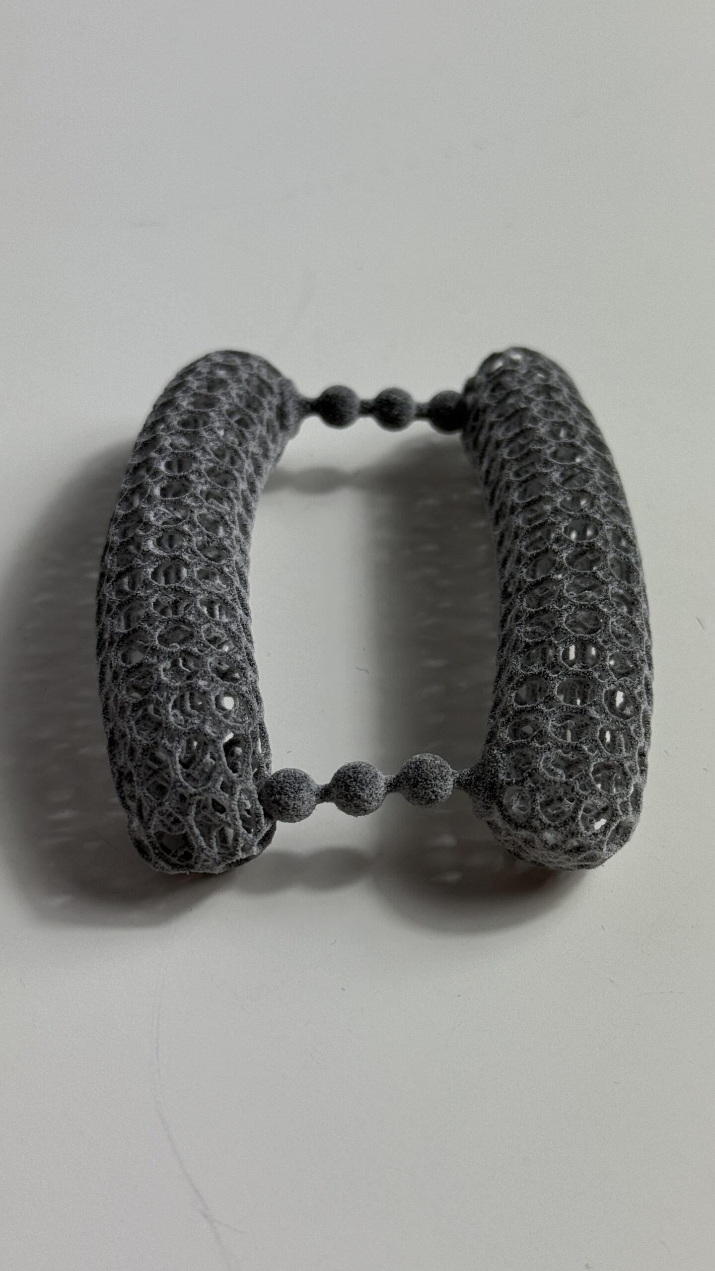
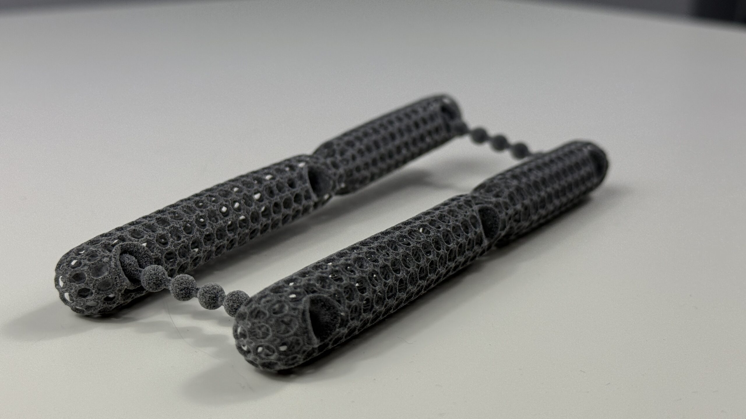
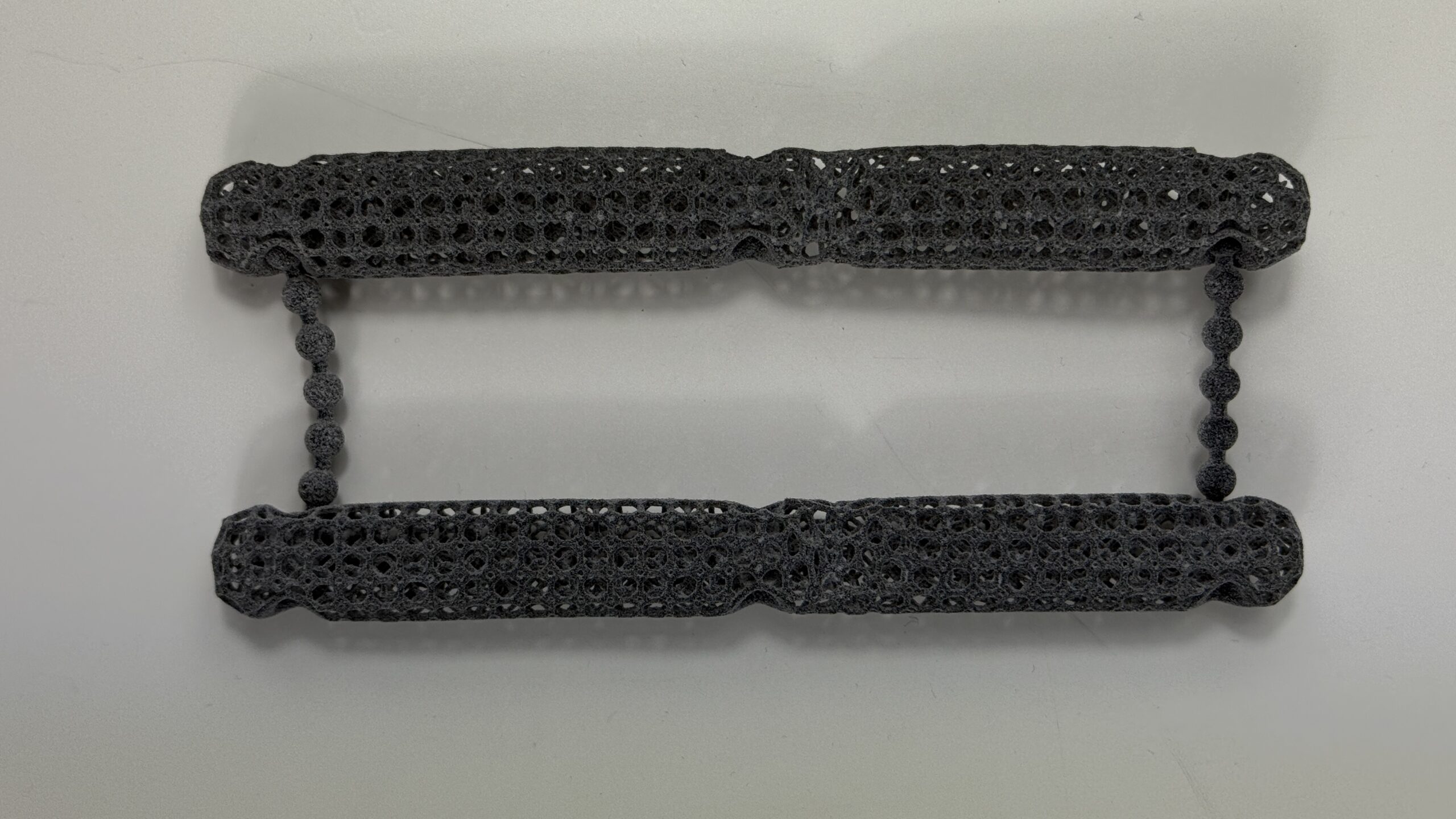
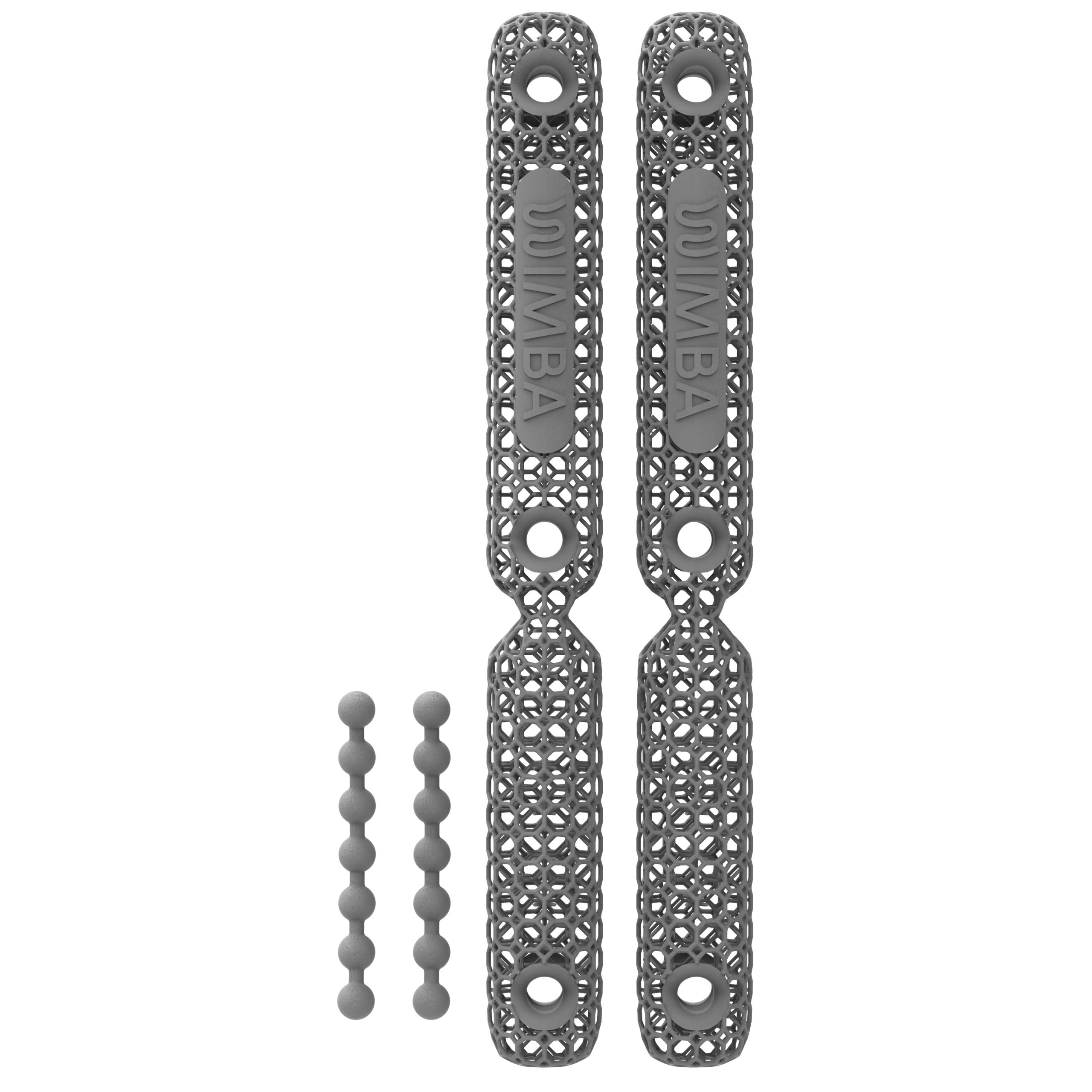
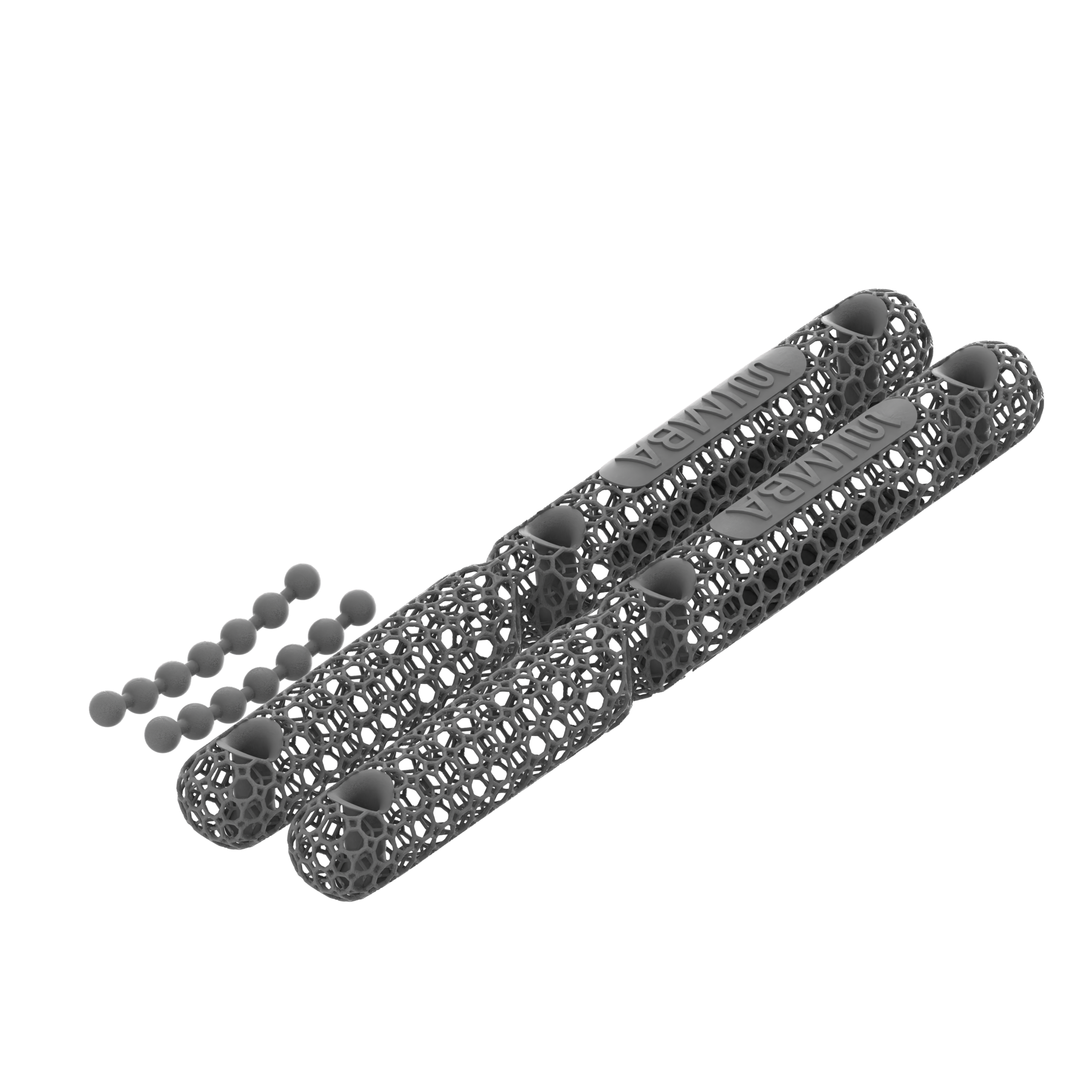
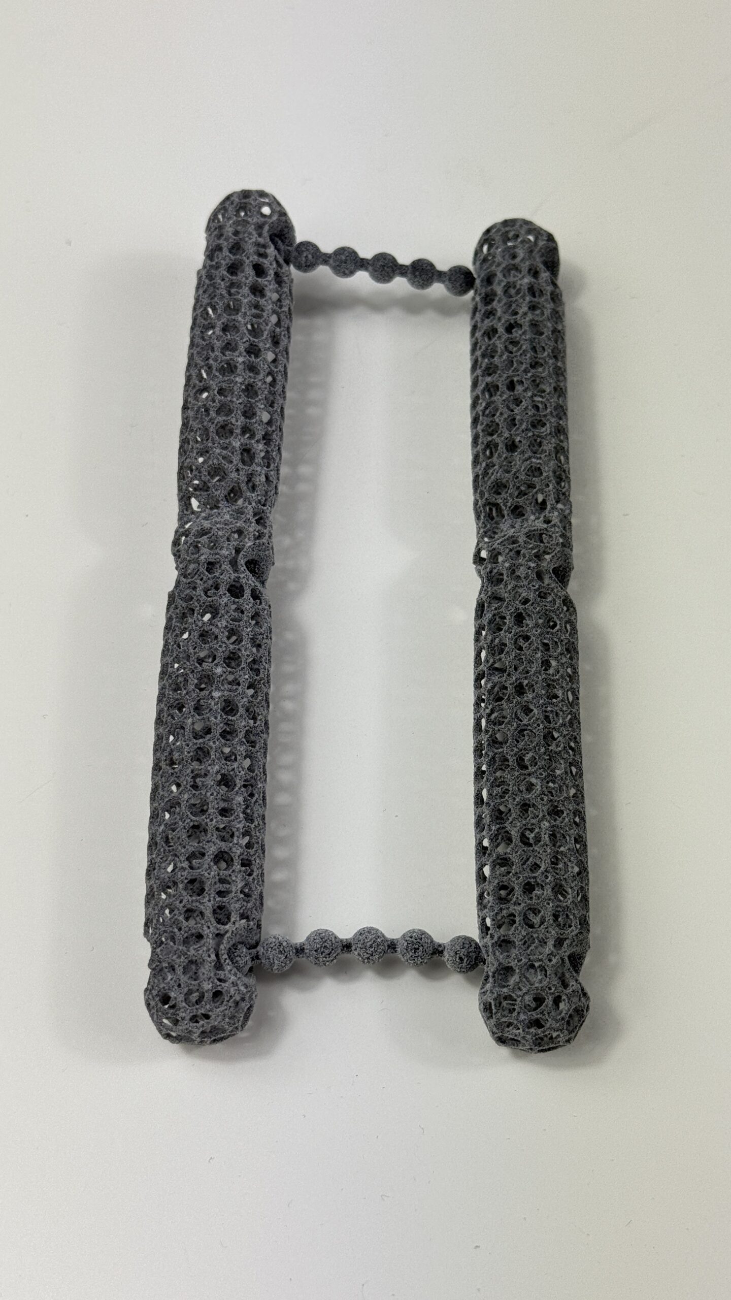
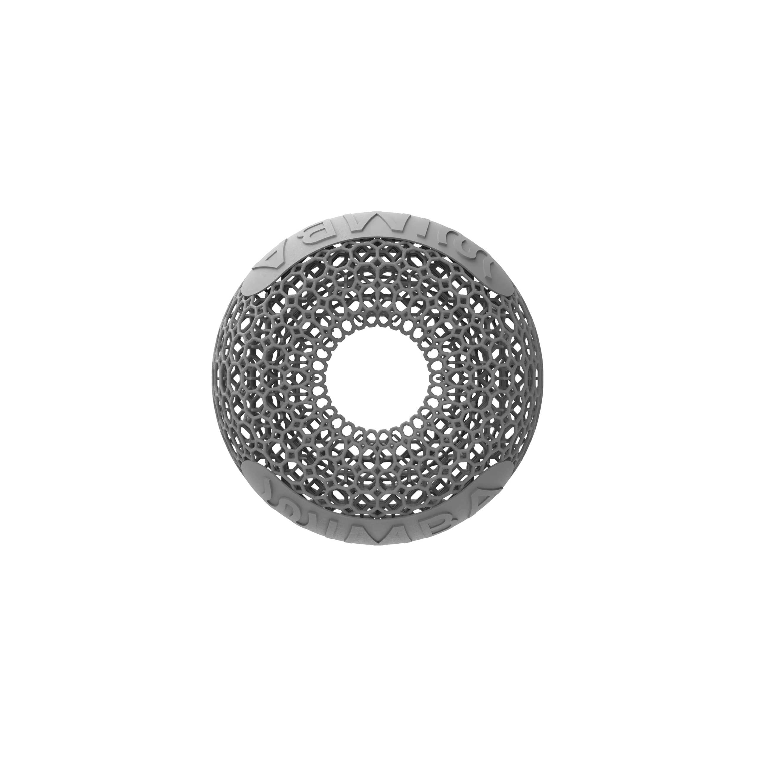
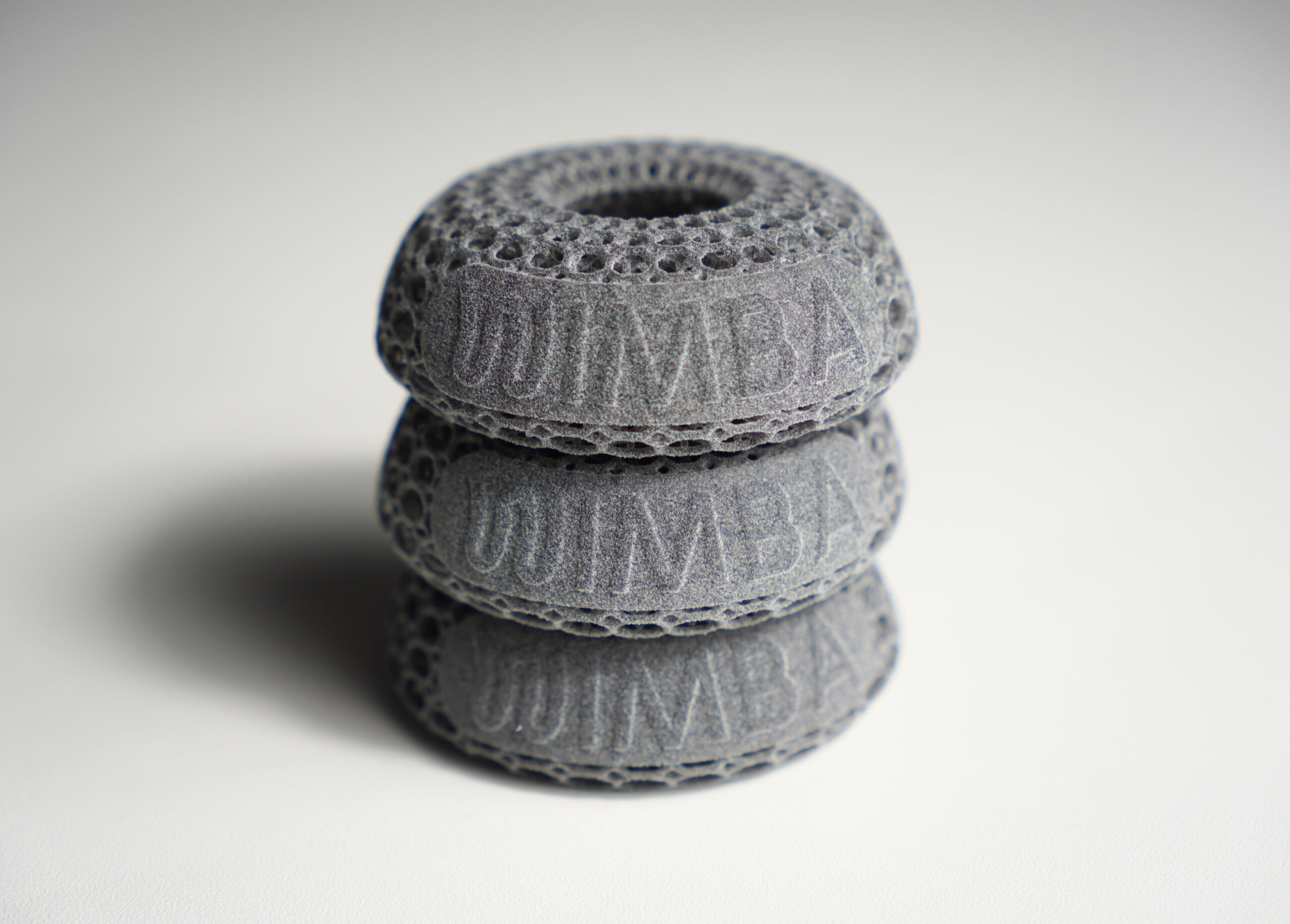
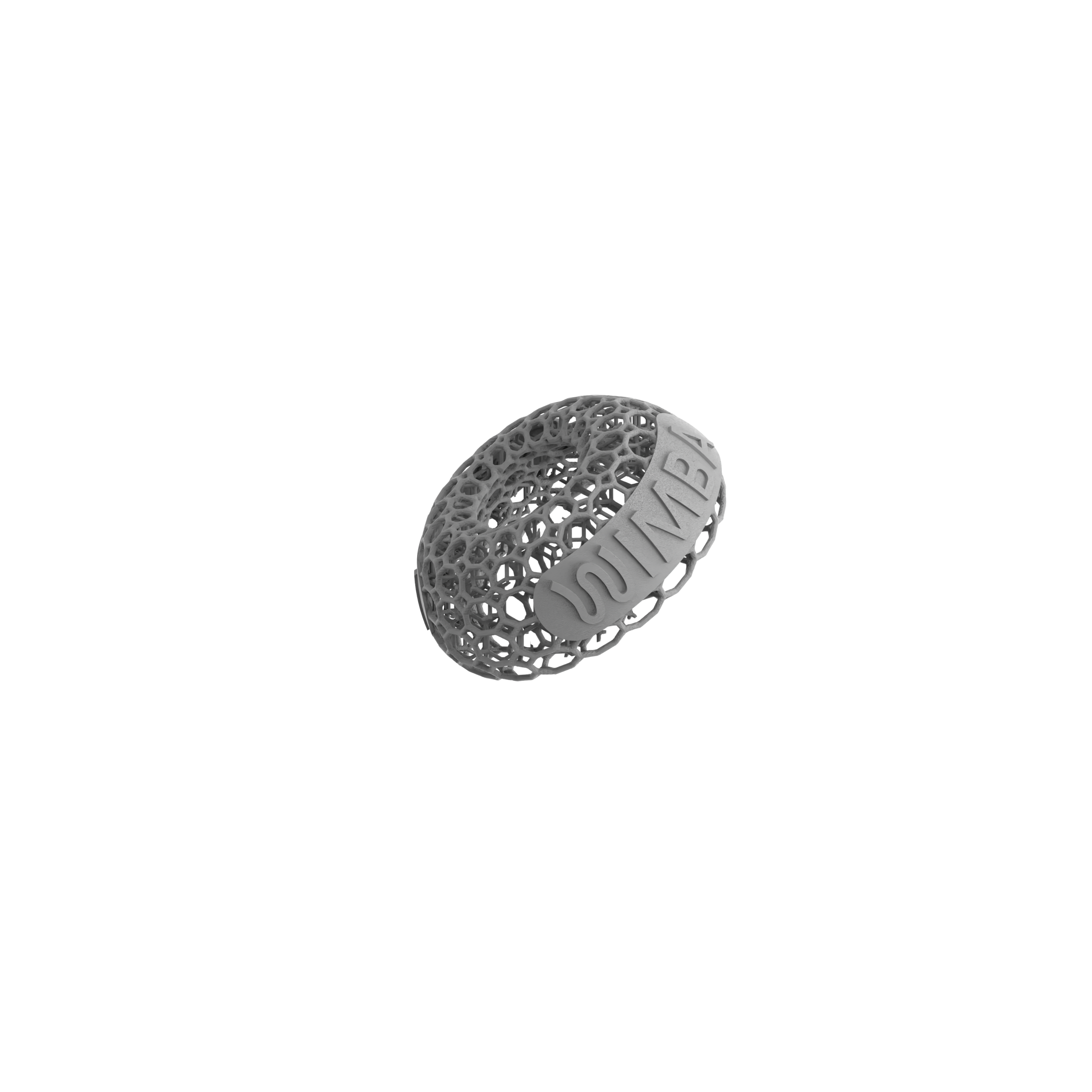
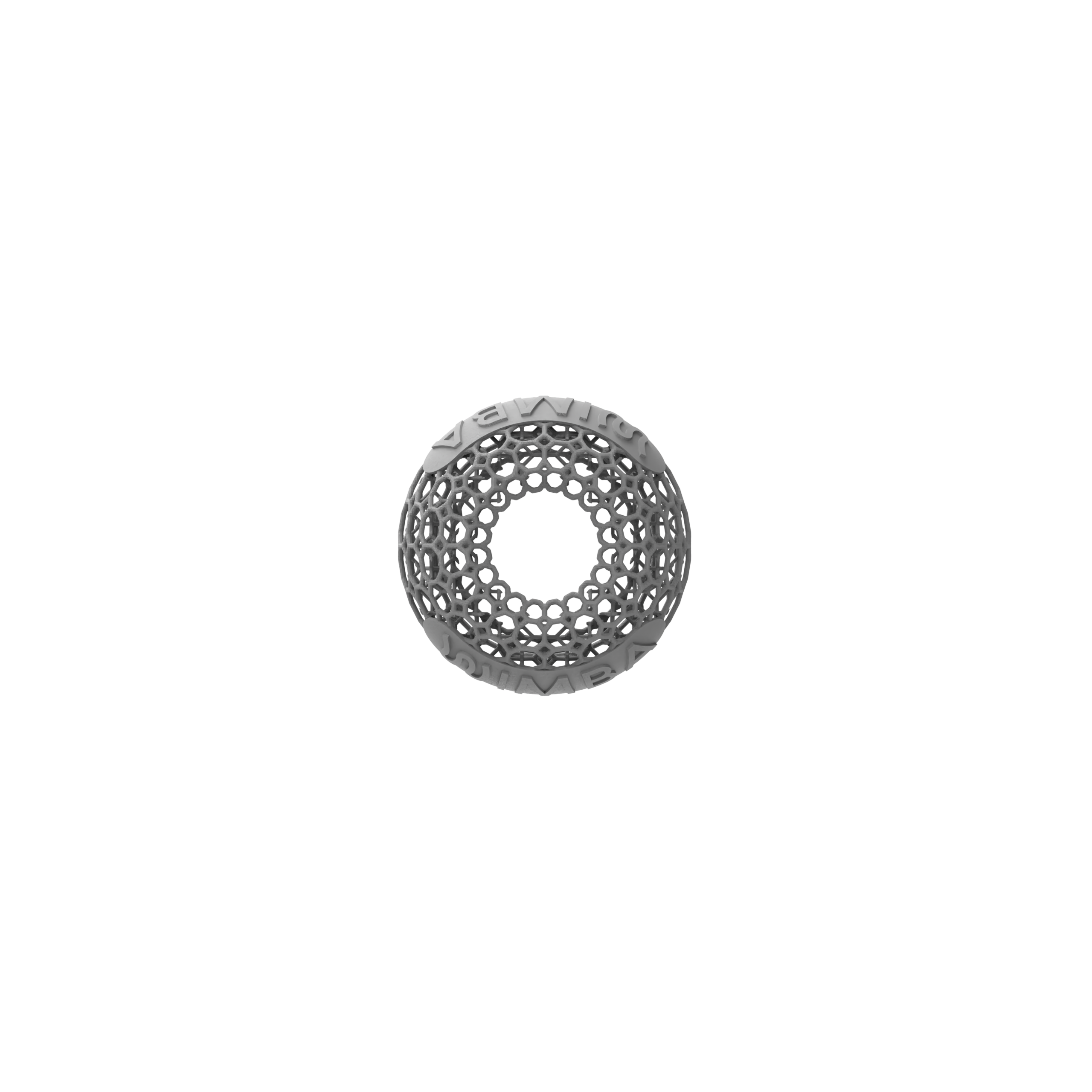
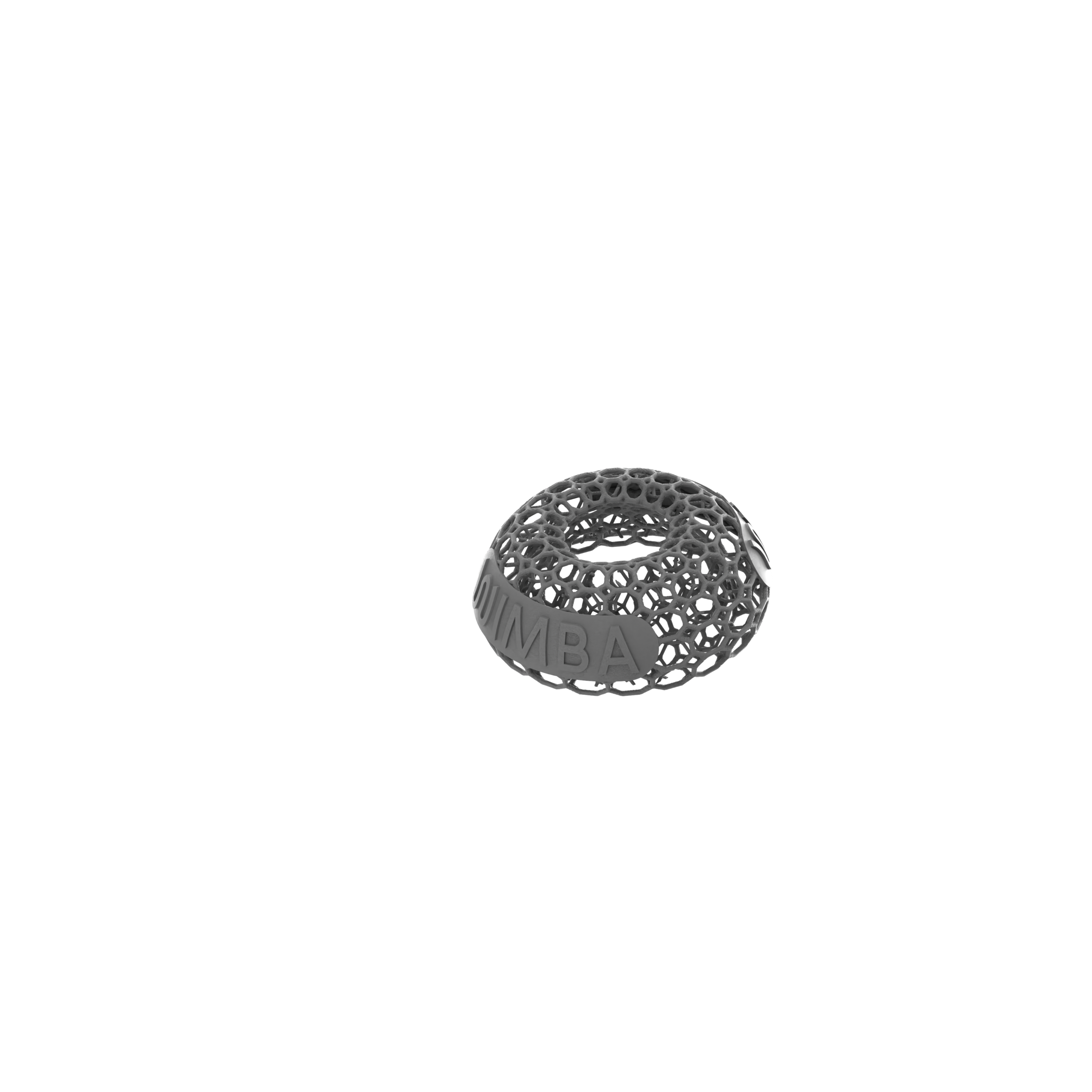
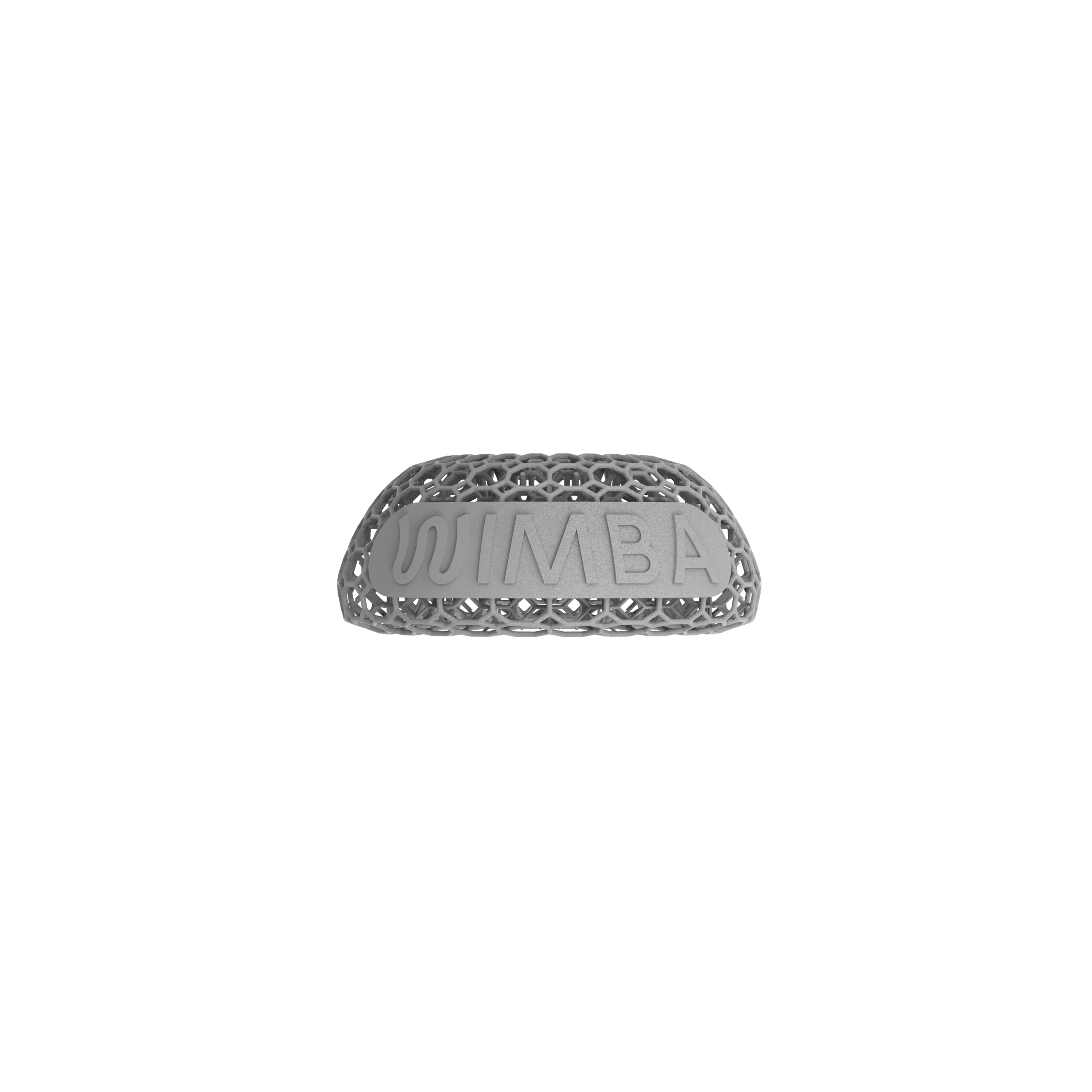
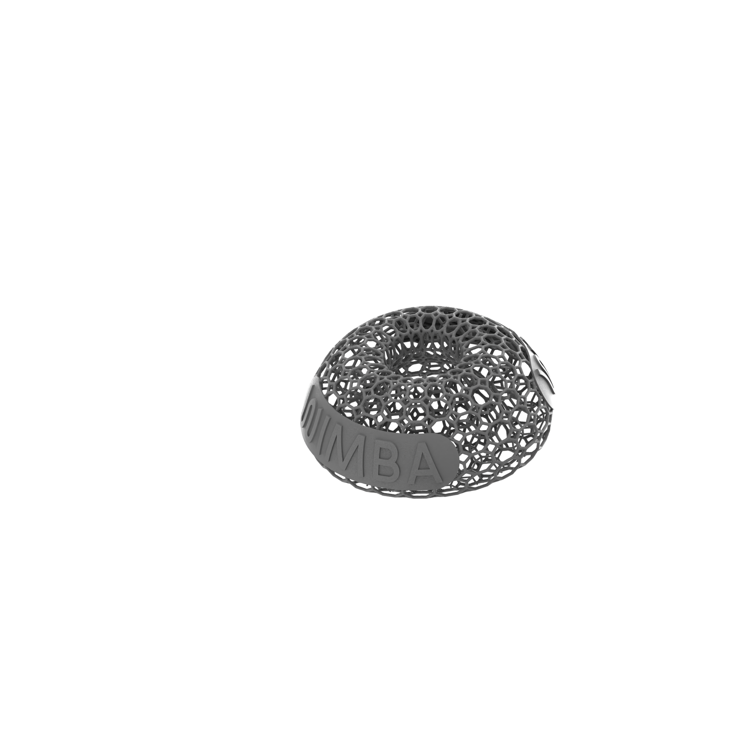
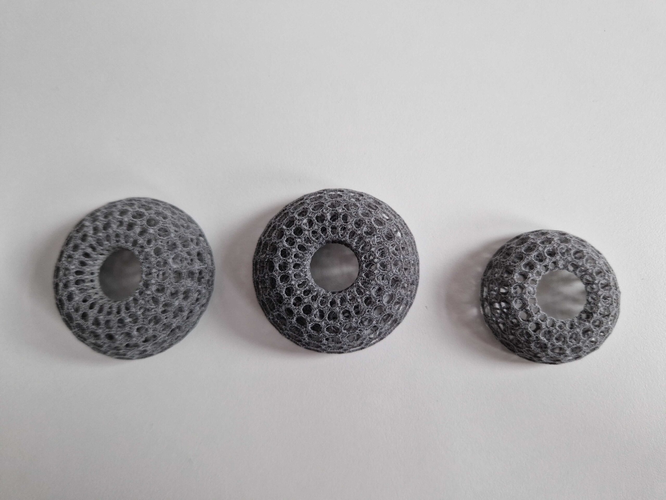
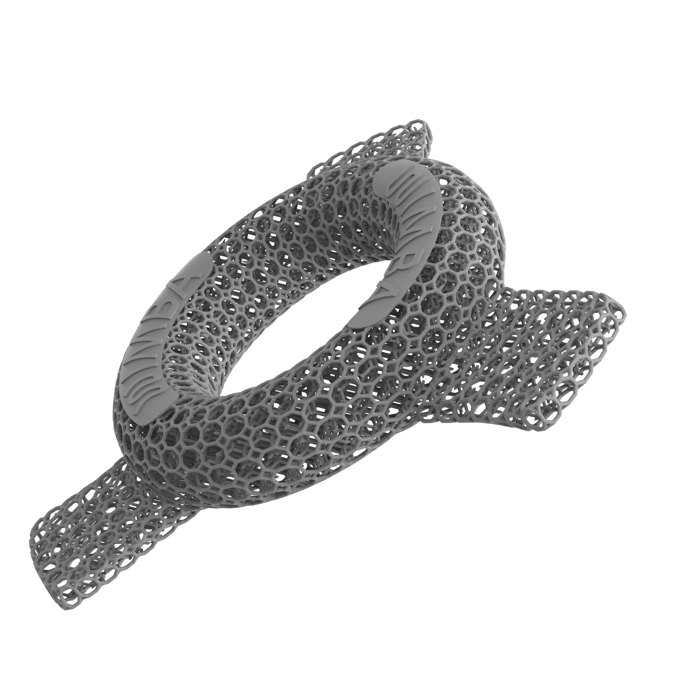
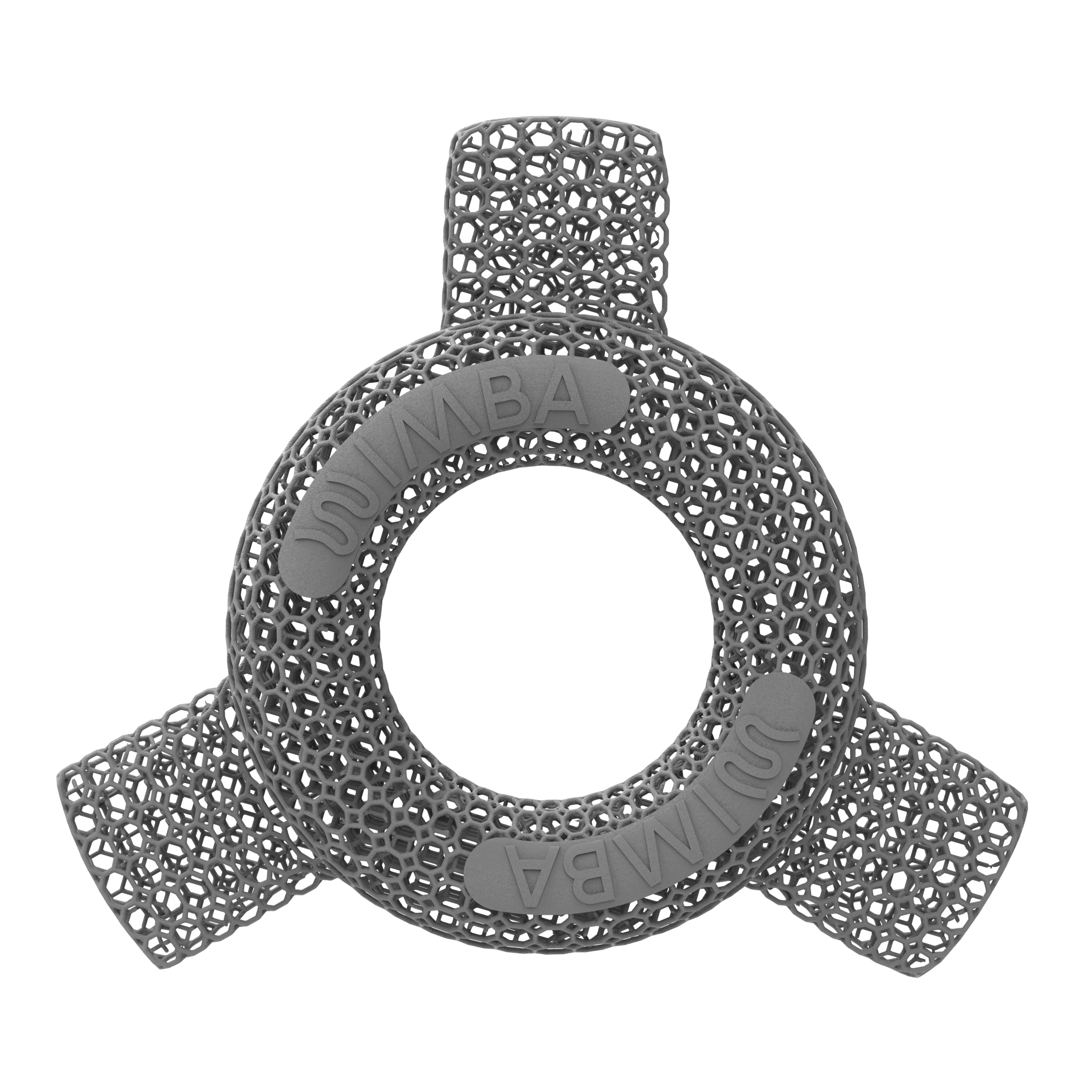
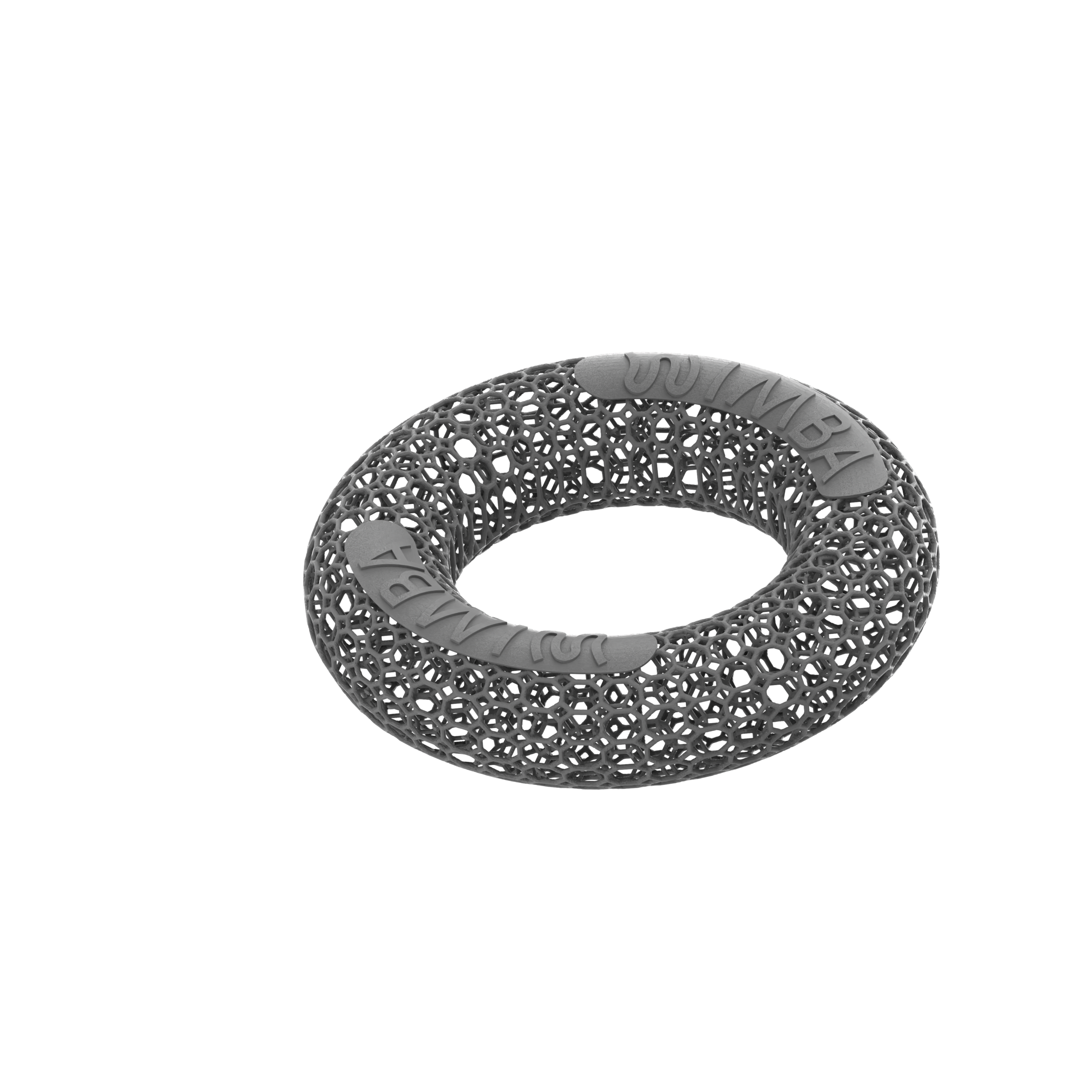
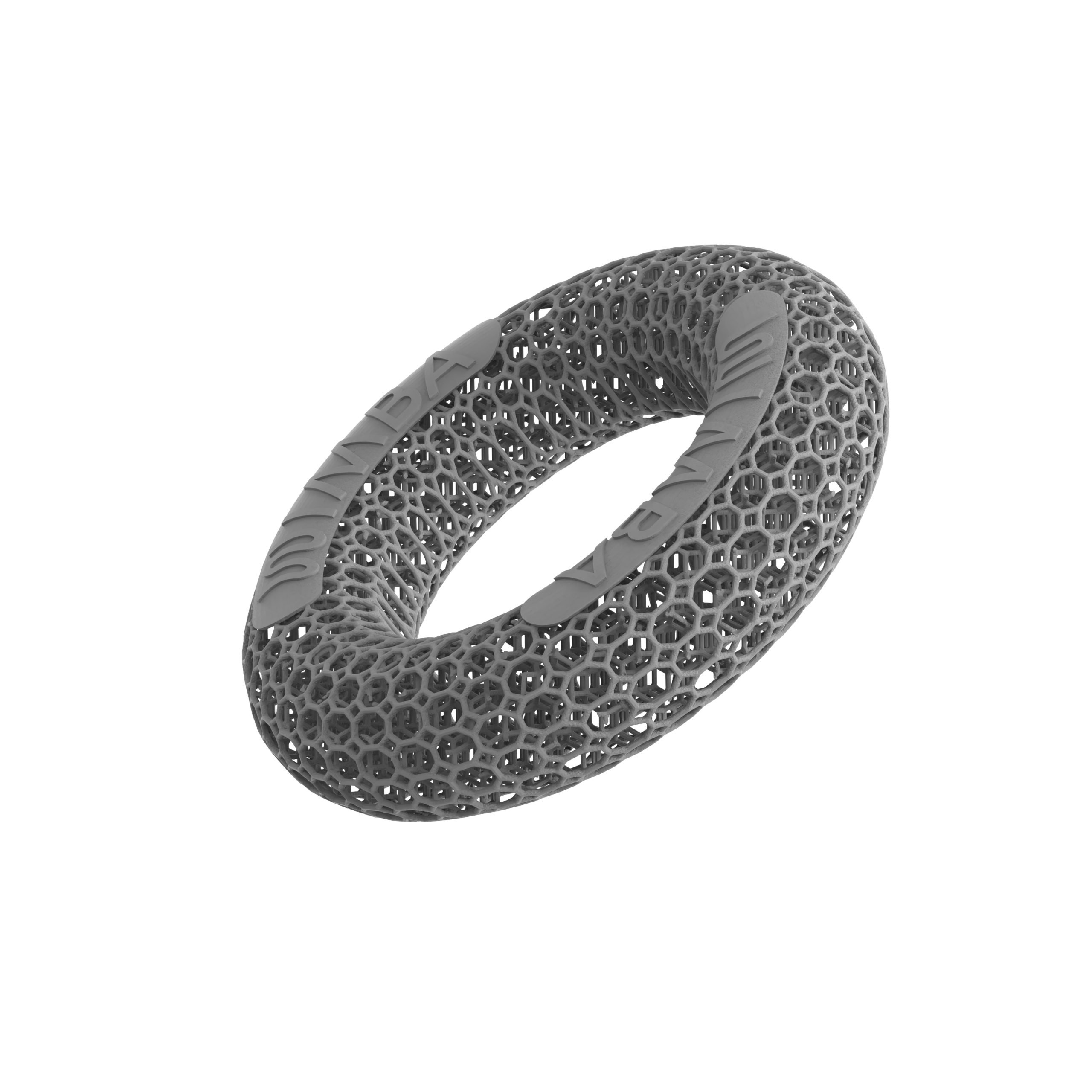
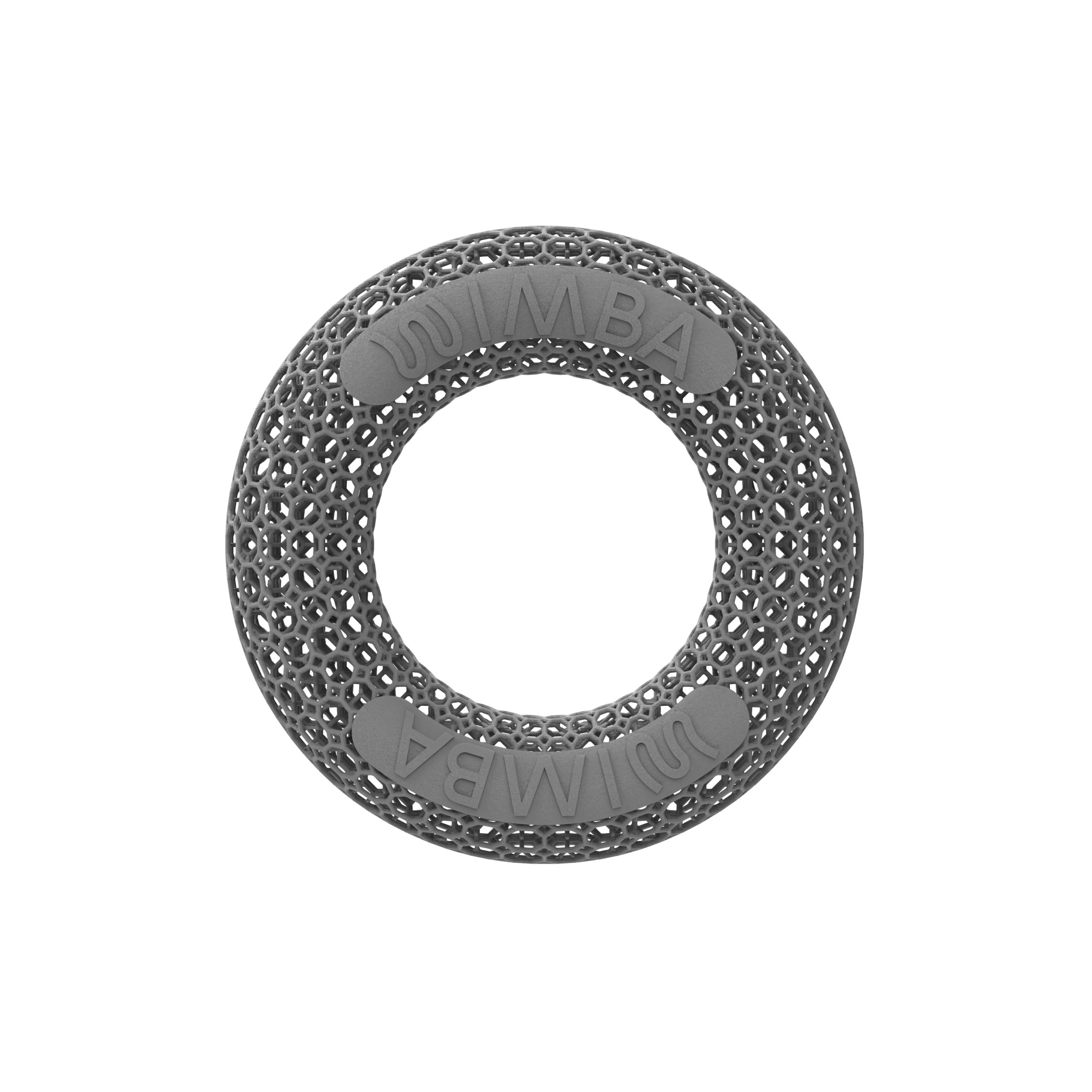
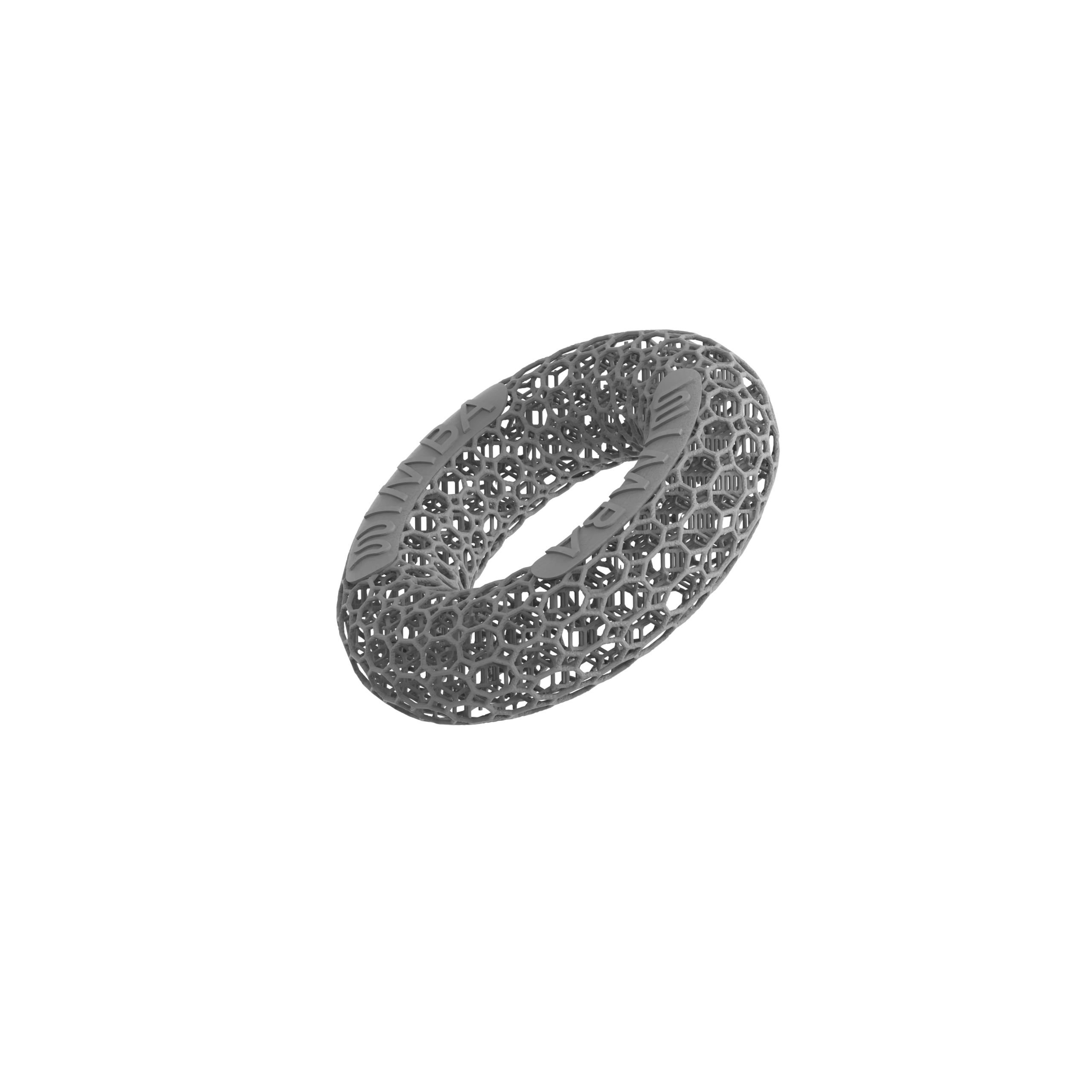
The WIMBA Cervical Collar GO is an adjustable, 3D-printed external support intended for selected C1–C2 cases in miniature and toy-breed dogs. It is available in five predefined sizes and is chosen from three patient measurements entered in WimbaAPP. Unlike WIMBA’s individually designed limb orthoses, the Cervical Collar GO is not produced from a 3D scan of the patient’s neck.
What the evidence actually shows
A retrospective study of 19 dogs treated nonsurgically for atlantoaxial subluxation reported a good final outcome in 10 of the 16 dogs with follow-up information.4 The protocol used historical cervical splints plus strict activity restriction and medical management; it did not evaluate the WIMBA device. The study supports the possibility of conservative care in selected patients, but it does not prove that any modern collar produces the same outcome or identify the best treatment for every case of cervical instability in dogs.
When surgery remains necessary
Surgical stabilization may be recommended when cervical instability in dogs is severe, neurological deficits are significant or progressive, or imaging shows a configuration unlikely to remain safe with external support alone. Published surgical series report neurological improvement in many treated dogs, while also documenting perioperative deaths, implant-related problems, and other complications.56 Those findings reinforce why treatment selection belongs with a veterinary neurologist or surgeon. A collar cannot replace stabilization or decompression when either is medically indicated.
Real WIMBA Patient Stories
The following stories show how external support has been used for cervical instability in dogs within three individual care plans. They are observational examples, not controlled evidence and not a promise of outcome for another affected dog.
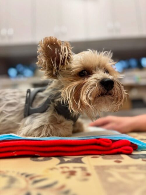
Kacperek: recovery after traumatic cervical injury
Kacperek, a 3.5-year-old Yorkshire Terrier, sustained severe traumatic cervical injury with spinal cord compression and tetraplegia. His conservative plan included intensive stabilization, pain management, monitoring, rehabilitation, and a WIMBA Cervical Collar GO. He later regained independent mobility. Because several interventions were used together, the outcome cannot be attributed to the collar alone.
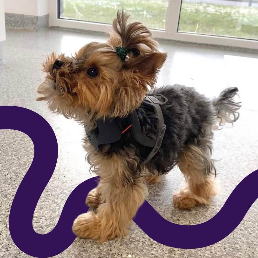
Pipi: complex coexisting conditions
Pipi’s case combined hydrocephalus with apex-rotational and upper-cervical instability. A lightweight WIMBA Cervical Collar GO was used under professional guidance as external support. The case demonstrates individualized planning; it does not mean that hydrocephalus alone causes cervical instability or makes every affected dog a collar candidate.

Coco: support for atlantoaxial instability
Coco was diagnosed with atlantoaxial instability and uses a WIMBA Cervical Collar GO within her care plan. Her story illustrates a selected congenital upper-cervical case in which external support was prescribed after diagnosis, rather than a brace being used as a substitute for veterinary assessment.
Who May Be a Candidate for a Cervical Collar?
When cervical instability in dogs is suspected, candidate selection begins with a confirmed diagnosis, neurological assessment, and appropriate imaging. According to WIMBA’s current product information, the Cervical Collar GO is intended for selected C1–C2 cases in miniature and toy-breed dogs. A veterinarian may consider it for selected atlantoaxial instability, other C1–C2 instability, selected C1–C2 fractures, or temporary support before or after surgery.
A collar may be considered when
- the diagnosis and unstable level have been established by a veterinarian;
- the clinical objective is clearly defined, such as limiting selected motion or providing temporary external support;
- the patient’s anatomy fits the available size range and safe adjustment is possible;
- the dog can be monitored closely for neurological change, fit, skin condition, and tolerance;
- the collar is integrated with activity restriction, medication, rehabilitation, surgery, or follow-up as prescribed.
A collar is not a first-aid shortcut
Do not use a cervical orthosis for dogs to manage an undiagnosed painful neck, acute collapse, worsening weakness, or suspected fracture. It is not a treatment for spinal cord compression and cannot make an unstable patient safe without veterinary stabilization. Severe or progressive cervical instability in dogs may require urgent surgery even when an external collar is available.
WIMBA Cervical Collar GO: Current Ordering Process
When the collar is prescribed for cervical instability in dogs, the ordering pathway differs from WIMBA’s scan-based custom orthoses. The WIMBA Cervical Collar GO uses five predefined sizes—Nano, XXS Short, XXS, XS, and S—and three patient measurements. No WimbaSCAN or WimbaBOX is required.
- Confirm the diagnosis and clinical objective. The prescribing team identifies the unstable region, neurological status, and purpose of support.
- Take three measurements. The veterinary provider follows the WIMBA guide and records the required patient dimensions.
- Enter the case in WimbaAPP. The measurements are used to select the closest of the five available collar sizes.
- Place the order. The adjustable collar is prepared and supplied through the veterinary workflow.
- Fit, introduce, and reassess. The team checks position, freedom from pressure, movement control, and neurological response, then sets the wear and follow-up schedule.
Fit must be monitored. Follow the prescribing team’s introduction schedule. Check the skin, padding, breathing, swallowing, behavior, and neurological function during use. Remove the collar and contact the veterinary team if it shifts, creates rubbing or pressure, changes breathing or swallowing, causes distress, or coincides with worsening pain, weakness, or coordination.
Frequently Asked Questions
What treatment options exist for dogs with cervical instability?
Cervical instability is not one diagnosis, so treatment depends on the affected level, underlying cause, imaging findings, spinal cord compression, and neurological status. A veterinarian-directed plan may include strict activity restriction, prescribed medication, carefully monitored external support, rehabilitation when appropriate, surgery, or a combination of these measures. WIMBA Cervical Collar GO may provide external support in selected C1–C2 cases or temporary support around surgery, but it does not replace surgical stabilization when surgery is indicated.
Can a cervical collar cure atlantoaxial instability in dogs?
No. A cervical collar provides external support but does not correct malformed bone, repair a ligament, or decompress the spinal cord. The veterinary team decides whether conservative management, surgery, or a combination is appropriate for the individual dog.
Is a dog neck brace a substitute for surgery?
No. When surgery is medically indicated, a dog neck brace cannot provide the internal stabilization or decompression the spinal cord may require. Treatment depends on the diagnosis, imaging, neurological status, progression, and the veterinary surgeon’s assessment.
How long should a dog wear a cervical collar?
There is no universal wear schedule. The prescribing veterinary team sets the duration and daily routine according to the diagnosis, treatment stage, skin health, tolerance, neurological response, and purpose of support. Follow-up may lead to adjustments or discontinuation.
Is hydrocephalus itself an indication for a cervical collar?
No. Hydrocephalus alone is not a standard indication for the WIMBA Cervical Collar GO. Pipi had hydrocephalus together with documented complex cervical instability, and her device was selected for her individual clinical situation under veterinary guidance.
How is the WIMBA Cervical Collar GO fitted?
A veterinary provider takes three patient measurements and enters them in WimbaAPP to select the closest of five predefined sizes. The collar is adjustable, but it is not individually designed from a 3D scan. The veterinary team checks and monitors the final fit.
What should I do if my dog suddenly becomes weak or painful in the neck?
Treat sudden neck pain, weakness, wobbliness, collapse, or inability to stand as an emergency. Minimize movement and contact a veterinarian or veterinary emergency service immediately. Do not test the neck or attempt to fit any external device before professional assessment.
Discuss Cervical Support With Your Veterinary Team
The next step for cervical instability in dogs is a diagnosis and a clearly defined treatment objective—not choosing a collar independently. If your veterinarian has identified a selected C1–C2 condition and is considering external support, review the current product information, submit the clinical case, or locate a WIMBA provider.
Medical disclaimer: This article is intended for general educational purposes only and is not a substitute for veterinary examination, diagnosis, or treatment. WIMBA devices are selected, fitted, and monitored by qualified veterinary professionals as part of an individualized clinical plan. Any dog showing neck pain, weakness, ataxia, collapse, or another neurological change should be evaluated promptly by a veterinarian or veterinary neurologist.
Scientific References
- Planchamp B, Forterre F, Vidondo B, et al. Determination of cutoff values on computed tomography and magnetic resonance imaging for the diagnosis of atlantoaxial instability in small-breed dogs. Veterinary Surgery. 2022;51(4):620–630. doi:10.1111/vsu.13799
- Hansen SC, Bacek LM, Kuo KW, Taylor AR. Traumatic atlantoaxial subluxation in dogs: eight cases (2009–2016). Journal of Veterinary Emergency and Critical Care. 2019;29(3):301–308. doi:10.1111/vec.12835
- Panya-ud C, Bunkrasin W, Keadwut K, et al. Correlation between outcomes of surgical correction of atlantoaxial instability in small dogs and magnetic resonance imaging abnormalities. Animals. 2026;16(9):1347. doi:10.3390/ani16091347
- Havig ME, Cornell KK, Hawthorne JC, McDonnell JJ, Selcer BA. Evaluation of nonsurgical treatment of atlantoaxial subluxation in dogs: 19 cases (1992–2001). Journal of the American Veterinary Medical Association. 2005;227(2):257–262. doi:10.2460/javma.2005.227.257
- Aikawa T, Shibata M, Fujita H. Modified ventral stabilization using positively threaded profile pins and polymethylmethacrylate for atlantoaxial instability in 49 dogs. Veterinary Surgery. 2013;42(6):683–692. doi:10.1111/j.1532-950X.2013.12016.x
- Stout Steele MW, Hodshon AW, Hopkins AL, et al. Multi-center retrospective evaluation of screw and polymethylmethacrylate constructs for atlantoaxial fixation in dogs. Veterinary Surgery. 2016;45(7):909–915. doi:10.1111/vsu.12533