 Skip to content

Atlantoaxial Instability in Dogs: Signs, Diagnosis, and Treatment
Atlantoaxial instability in dogs is an uncommon but potentially life-threatening disorder of the joint between the first two cervical vertebrae, the atlas (C1) and axis (C2). Abnormal motion at this level can injure or compress the upper cervical spinal cord. Clinical signs range from neck pain to incoordination, weakness, paralysis, and, in the most severe cases, respiratory arrest.12
This evidence-based guide explains what atlantoaxial instability in dogs means, why appearance alone cannot define its cause or severity, how veterinarians investigate it, and what published outcome studies say about conservative and surgical management. It also explains where a dog neck brace for AAI or other cervical support for dogs may fit without presenting external support as a substitute for diagnosis or internal surgical stabilization.
The short answer
Suspected atlantoaxial instability in dogs is not a condition to test at home. Do not flex, rotate, massage, or repeatedly examine a painful neck. Keep movement to the minimum needed for safe transport and seek prompt veterinary care. A neurological examination and carefully planned imaging are used to assess the C1-C2 region, spinal cord, and possible concurrent craniocervical abnormalities. Treatment is individualized; some selected dogs are managed with strict activity restriction and external support, while many dogs with neurological deficits, persistent pain, marked displacement, or failed conservative care are considered for surgery.12
What Atlantoaxial Instability in Dogs Actually Means
Understanding atlantoaxial instability in dogs begins with the normal joint. The atlas supports the skull, while the axis provides a pivot around which the atlas and head can rotate. The dens, a projection from C2, works with the transverse, alar, apical, dorsal, and ventral supporting structures to maintain alignment. Cadaveric research shows that these ligaments have distinct structural roles and that combined flexion and rotation places substantial strain on the alar ligaments.4
With canine atlantoaxial instability, the stabilizing system is insufficient and C1 and C2 move abnormally relative to one another. The axis may move dorsally toward the vertebral canal, producing dynamic or persistent spinal cord compression. The term atlantoaxial subluxation in dogs describes the resulting partial loss of normal alignment; instability refers to the abnormal motion that permits or accompanies it. When discussing atlantoaxial instability in dogs, the terms are often used together in veterinary publications, but they do not describe every patient’s anatomy in exactly the same way.12
| Term | What it describes | What it does not establish alone |
|---|---|---|
| Atlantoaxial instability | Abnormal motion or inadequate stability between C1 and C2 | The underlying congenital or traumatic mechanism |
| Atlantoaxial subluxation | Partial loss of normal C1-C2 alignment | The amount of spinal cord injury or the best treatment |
| Cervical myelopathy | Neurological dysfunction caused by disease affecting the cervical spinal cord | That AAI is the cause without localization and imaging |
| Dens abnormality | Aplasia, hypoplasia, separation, angulation, fracture, or another alteration of the odontoid process | That the dens is the only deficient stabilizing structure |
This distinction matters because atlantoaxial instability in dogs is not simply a missing dens. Ligamentous insufficiency, incomplete ossification of the atlas, abnormal dens morphology, atlanto-occipital overlapping, other craniocervical malformations, fracture, and traumatic ligament disruption may occur alone or in combination.356
What Causes Atlantoaxial Instability in Dogs?
Congenital or developmental atlantoaxial instability in dogs is reported most often in young toy and small-breed patients. Reviews consistently identify Yorkshire Terriers, Chihuahuas, Pomeranians, Miniature and Toy Poodles, and Pekingese among the frequently represented breeds, although other breeds, larger dogs, and cats can be affected.12
The phrase “congenital AAI” does not mean every affected puppy is born with visible spinal cord signs. A predisposing malformation or ligamentous weakness may exist before symptoms appear, and a seemingly minor incident may trigger acute decompensation. Conversely, truly traumatic atlantoaxial instability in dogs can follow fracture or ligament disruption in an animal without a known developmental abnormality and can occur at any age or body size.13
AAI may be part of a more complex craniocervical problem
A multicenter case-control study found that small-breed dogs with AAI were more likely than controls to have other craniovertebral junction abnormalities, including atlanto-occipital overlapping and dens anomalies.6 A later retrospective study of 41 toy-breed dogs also documented multiple concurrent imaging findings.7 This is one reason a treatment plan cannot be based only on breed, age, or one radiographic distance.
In a surgical cohort of 106 dogs with canine atlantoaxial instability, 70 had dens abnormalities and 75 had incomplete ossification affecting at least part of the dorsal neural arch of C1.5 These findings show that both abnormalities are common in selected referral populations, but neither was present in every dog and the cohort cannot establish population prevalence.
Signs of Atlantoaxial Instability in Dogs
The clinical picture of atlantoaxial instability in dogs depends on whether abnormal movement causes pain alone, spinal cord compression, concussion, or more severe injury. Signs may be progressive, intermittent, or sudden, including after minor trauma in a predisposed dog.12
- Neck pain, vocalization, a guarded posture, or reluctance to move the head
- Low head carriage or difficulty lowering the head to eat or drink
- Ataxia, inconsistent paw placement, crossing limbs, or loss of balance
- Weakness affecting two or all four limbs
- Inability to stand or walk, or complete paralysis
- Rapid deterioration after a fall, jump, collision, or forceful neck movement
- In the most severe upper-cervical injuries, impaired breathing, collapse, or death
These signs are not specific to atlantoaxial instability in dogs. Intervertebral disc disease, meningomyelitis, fracture, other craniocervical abnormalities, spinal cord tumors, and several painful or neurological disorders can produce overlapping signs. A painful small dog should therefore not be placed into a neck brace merely because AAI seems plausible.
Seek emergency veterinary care
Treat inability to stand, rapidly worsening weakness, severe pain, collapse, altered consciousness, or difficulty breathing as an emergency. Minimize neck movement during transport and contact the receiving clinic for handling instructions. Do not test whether the dog can walk, repeatedly lift the head, or attempt to apply a rigid homemade support.
How Atlantoaxial Instability in Dogs Is Diagnosed
Diagnosing atlantoaxial instability in dogs begins by localizing the problem through history, general examination, and neurological examination. The clinician considers the onset, trauma history, pain, gait, postural reactions, limb strength, breathing, and whether signs localize to the upper cervical spinal cord. Imaging then evaluates alignment, osseous anatomy, soft tissues, spinal cord compression, and possible concurrent abnormalities.13
| Diagnostic component | What it may show | Important limitation |
|---|---|---|
| Neurological examination | Pain, gait abnormality, postural deficits, weakness, and neuroanatomic localization | It cannot show the C1-C2 anatomy or identify every cause |
| Neutral radiographs | Alignment, C1-C2 overlap, angulation, dens morphology, fracture, and other bony changes | Some measurements may remain normal in individual affected dogs |
| Flexed radiographs | Dynamic separation or angulation under a standardized protocol | Positioning alters measurements and must be selected and performed by the clinical team |
| Computed tomography | Detailed bony morphology, joint relationships, measurements, and surgical corridors | Bone detail alone does not quantify every aspect of spinal cord injury |
| Magnetic resonance imaging | Spinal cord compression and signal change, ligaments and other soft tissues, and concurrent neurological disease | Interpretation still requires clinical correlation and appropriate positioning |
Neutral radiographs can provide objective evidence. In a study involving 92 control toy-breed dogs and 10 dogs with AAI, reduced overlap between C1 and C2 was the most sensitive and specific measurement assessed, although several other indices had lower sensitivity and could be normal in individual cases.8 This supports measurement-led interpretation rather than a single visual impression.
Flexed radiographs have also been studied. A multicenter investigation established diagnostic cutoffs using a standardized degree of flexion in sedated or anesthetized toy-breed dogs.9 That finding does not make unsupervised neck flexion safe. Position, sedation or anesthesia, airway management, and whether a dynamic view is justified are clinical decisions for the imaging team.
CT and MRI add different information. A 2022 multicenter study established several cross-sectional imaging cutoffs for small-breed dogs and emphasized that the decision for surgery should still combine clinical and imaging findings rather than follow one number.10 Advanced imaging is particularly valuable when atlantoaxial instability in dogs is anatomically complex, when surgery is being planned, or when another craniocervical or spinal cord disorder may coexist.
Conservative or Surgical Treatment for Atlantoaxial Instability in Dogs?
There is no randomized trial showing that one treatment pathway for atlantoaxial instability in dogs is best for every patient. Published evidence consists mainly of retrospective case series, surgical technique reports, anatomical studies, imaging studies, and expert reviews. Treatment selection must consider neurological status, duration and progression of signs, degree and direction of displacement, fractures or malformations, spinal cord injury, concurrent conditions, anesthetic risk, patient size, and the owner’s ability to manage intensive aftercare.12
What conservative management usually involves
Published nonsurgical management of atlantoaxial instability in dogs combines external cervical coaptation with strict activity restriction and prescribed medical care. Its objective is to limit motion while fibrous stabilization develops and the spinal cord is protected from additional movement. This is not the same as permanently fusing C1 and C2, and recurrence or deterioration remains possible after external support is removed.111
The most frequently cited nonsurgical outcome study of atlantoaxial subluxation in dogs reviewed 19 patients treated using a cervical splint. A good final outcome was reported for 10 of the 16 dogs with evaluable final outcomes. Dogs with clinical signs lasting 30 days or less were more likely to have a good long-term result. The authors considered splint management viable particularly for young dogs experiencing a first acute episode.11
That result is encouraging but limited: the study was retrospective, small, had no surgical or untreated control group, and does not show that all mild cases will recover or that dogs with major deficits should routinely avoid surgery. It also evaluated historical splint constructs, not WIMBA Cervical Collar GO.
What surgical stabilization aims to do
Surgery aims to realign and rigidly stabilize the atlantoaxial joint, often promoting fusion. Reviews generally favor surgical management for dogs with neurological deficits, persistent or severe pain, clear instability, progression, or failure of conservative treatment, while recognizing that the anatomy is technically demanding and complications can be serious.12
In a retrospective single-surgeon series of 49 dogs undergoing modified ventral stabilization, two dogs died during the perioperative period. Among 47 dogs with follow-up, 46 improved neurologically; partial pin breakage occurred in 16 dogs without reported neurological deterioration.12 These results describe one technique in a selected referral population and should not be treated as a universal success rate for surgery.
| Evidence set | Reported finding | What it cannot prove |
|---|---|---|
| Nonsurgical splint series 19 dogs | Good final outcome in 10 of 16 dogs with evaluable final outcomes; shorter duration of signs was associated with a better result.11 | That a brace is equivalent to surgery or suitable for every neurological grade |
| Ventral surgical series 49 dogs | 46 of 47 dogs with follow-up improved neurologically; two perioperative deaths and partial pin breakage in 16 dogs were reported.12 | That all techniques, surgeons, anatomical patterns, or patients have the same outcome |
The evidence does not support a simple severity rule
It is too simplistic to write that “mild and moderate AAI can often be treated without surgery” while “severe AAI always needs surgery.” The small nonsurgical series included varying neurological grades, and imaging anatomy, progression, duration, concurrent abnormalities, and patient safety all influence selection. A veterinary neurologist or surgeon should interpret atlantoaxial instability in dogs as an individual clinical and imaging problem.
When a Dog Neck Brace for AAI May Be Considered
For atlantoaxial instability in dogs, a dog neck brace for AAI is an external support, not an internal fixation construct. Its intended role is to help limit unwanted cervical movement and maintain the fitted position as one part of a veterinarian-directed plan. It does not remove a malformed dens, repair a torn ligament, decompress the spinal cord, create guaranteed C1-C2 fusion, or eliminate the possibility of sudden deterioration.
External cervical support for dogs may be considered in selected conservative cases, as temporary support before surgery, or during postoperative care when the treating specialist specifically includes it. The exact position, duration, wearing schedule, activity restriction, medication, and recheck plan must follow the diagnosis and surgical strategy rather than a generic protocol.
Because a dog neck brace for AAI sits close to the airway, mouth, ears, eyes, and skin, fitting and monitoring are clinically important. The patient must be able to breathe and swallow normally, the collar must remain in the intended position, and contact areas require regular checks. Any change in breathing, swallowing, comfort, neurological function, device position, or skin condition needs prompt veterinary review.
What published brace evidence can and cannot tell us
The 2005 nonsurgical series supports the possibility of favorable outcomes in selected dogs managed with a cervical splint, but it does not identify one ideal brace design or prove complete immobilization.11 The peer-reviewed studies cited in this article did not evaluate WIMBA Cervical Collar GO. Outcomes from historical splints therefore cannot be transferred directly to this product.
Where WIMBA Cervical Support for Dogs Fits
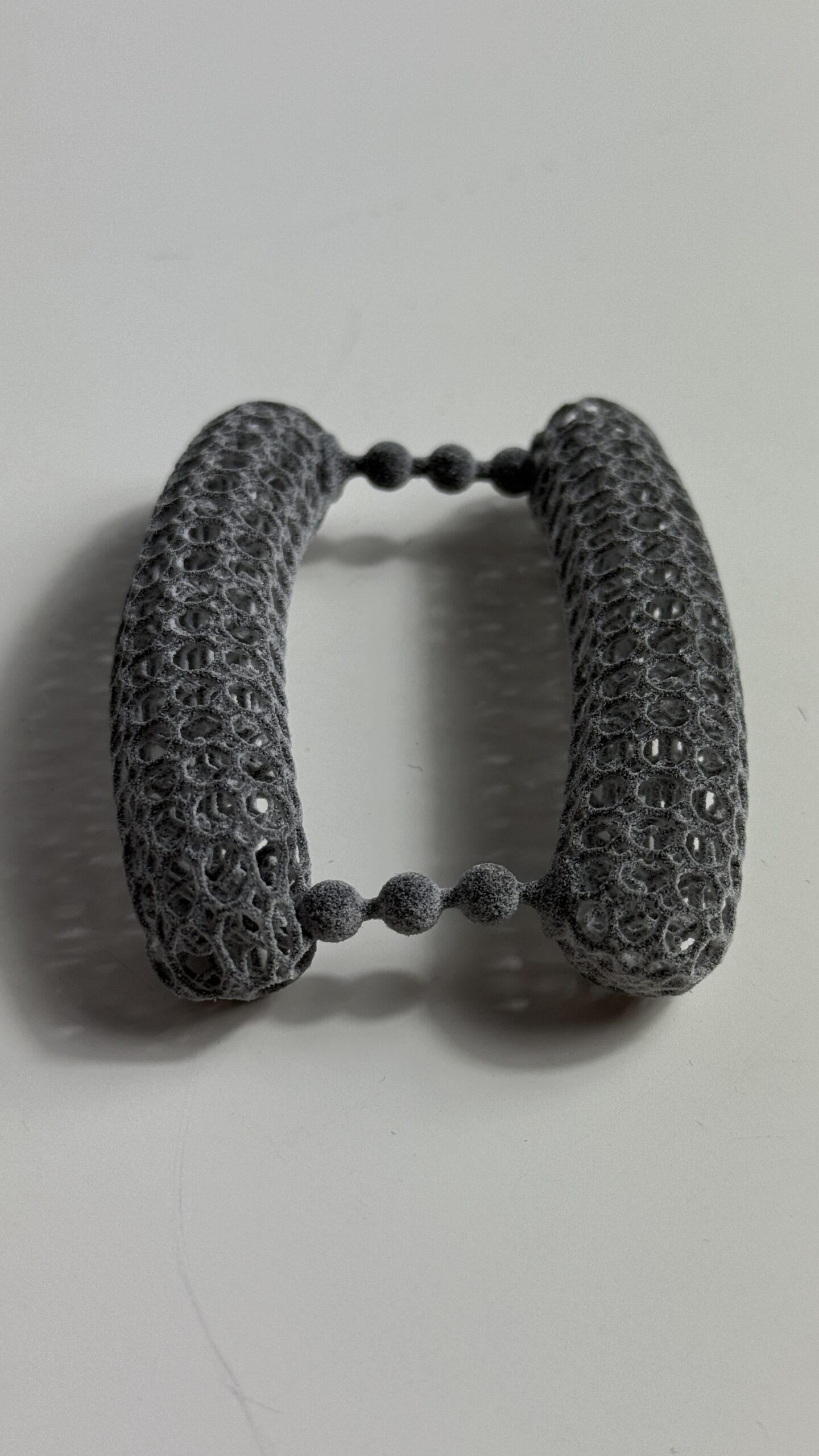
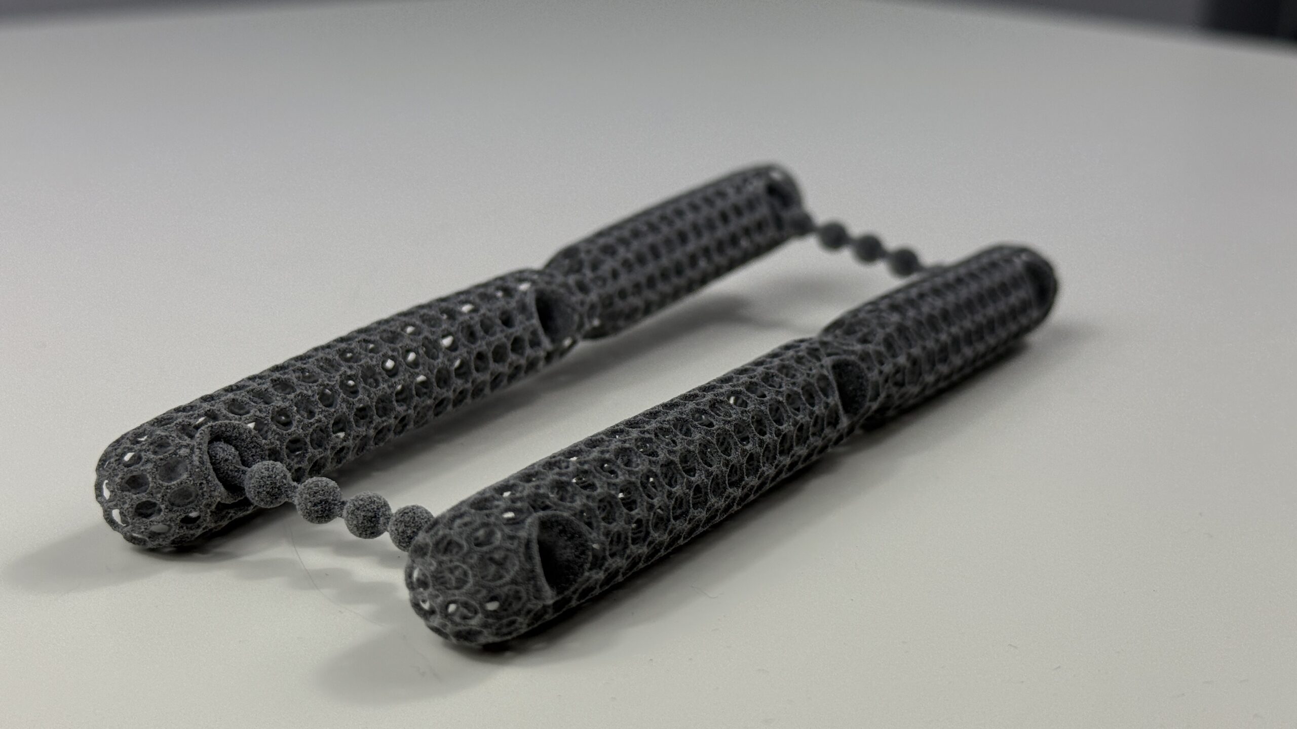
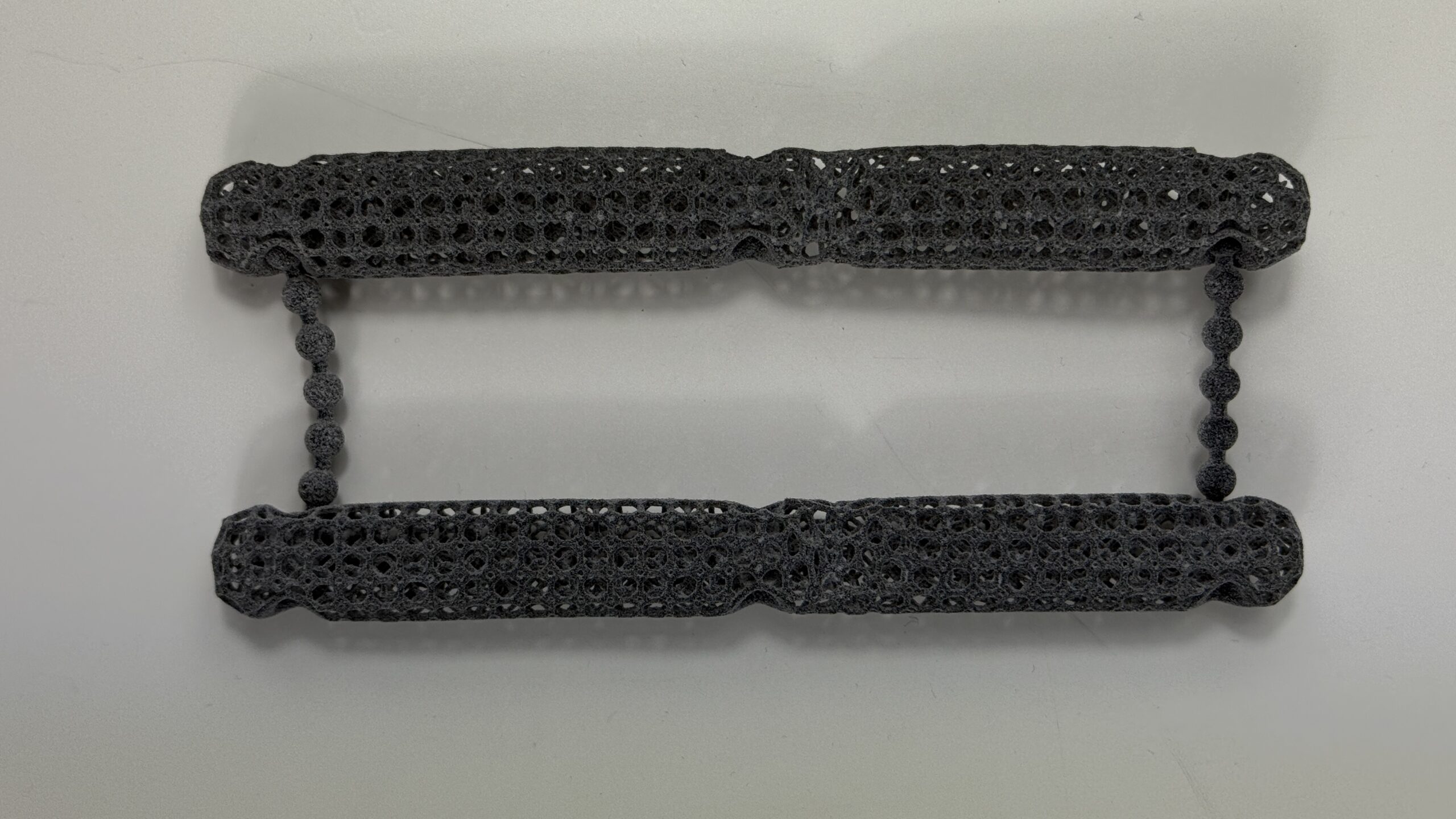
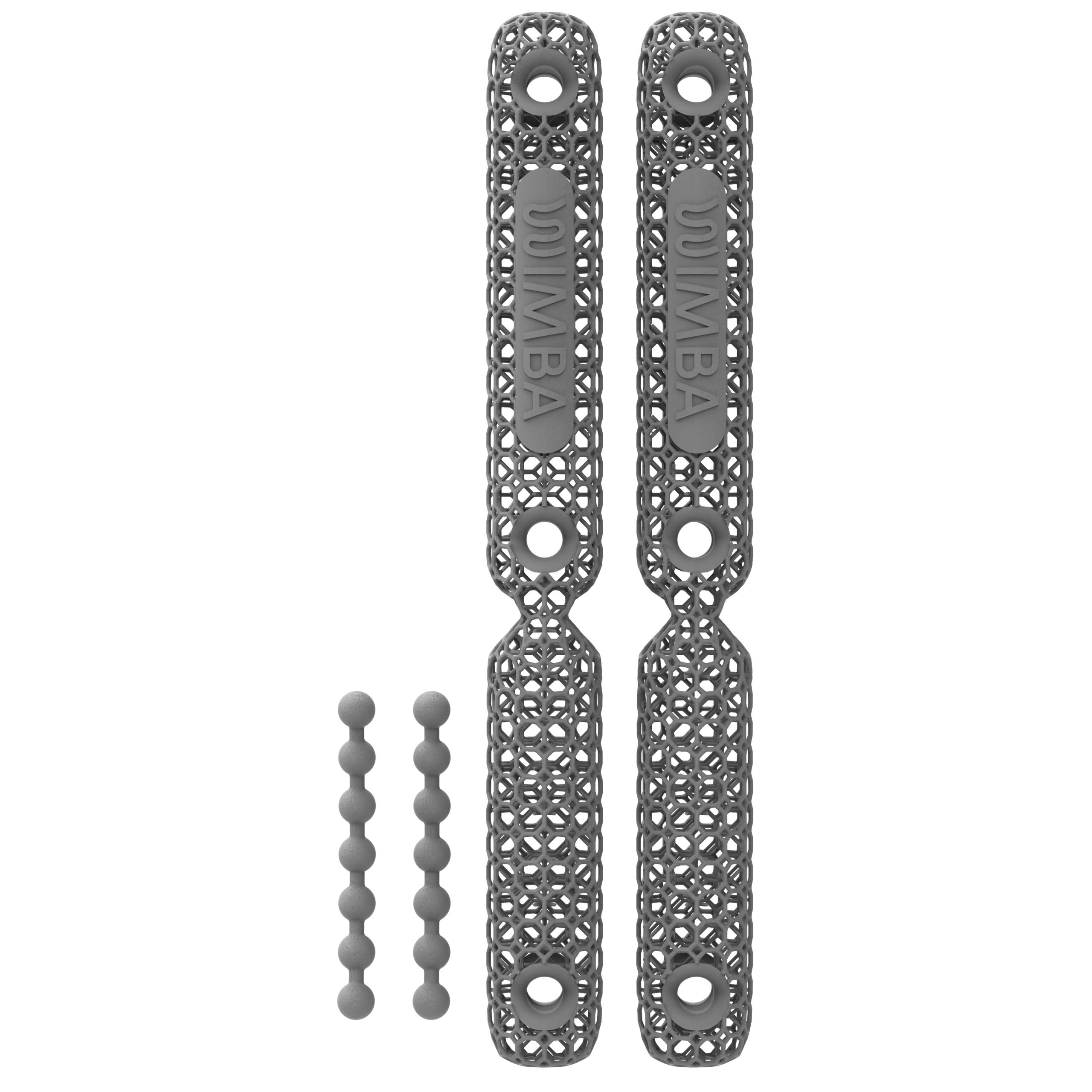
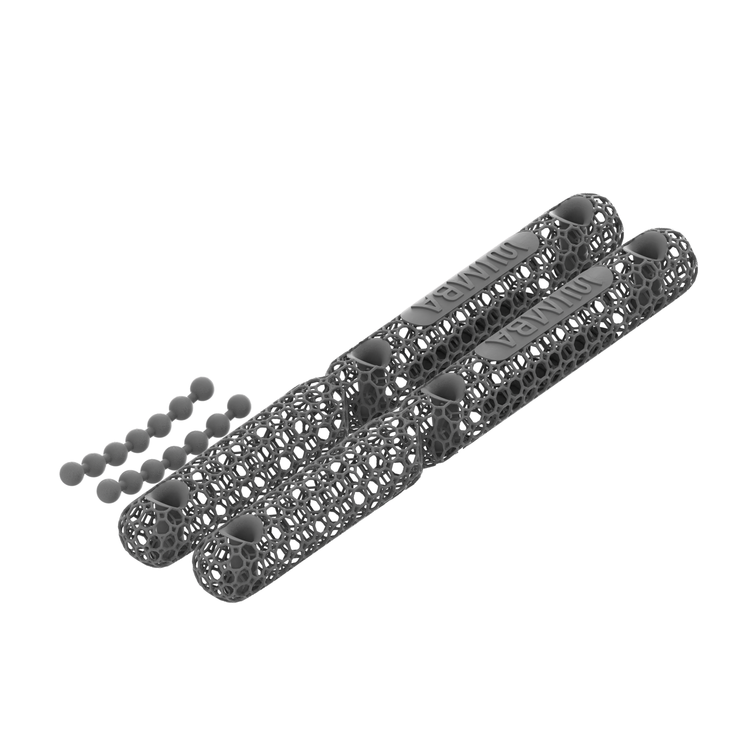
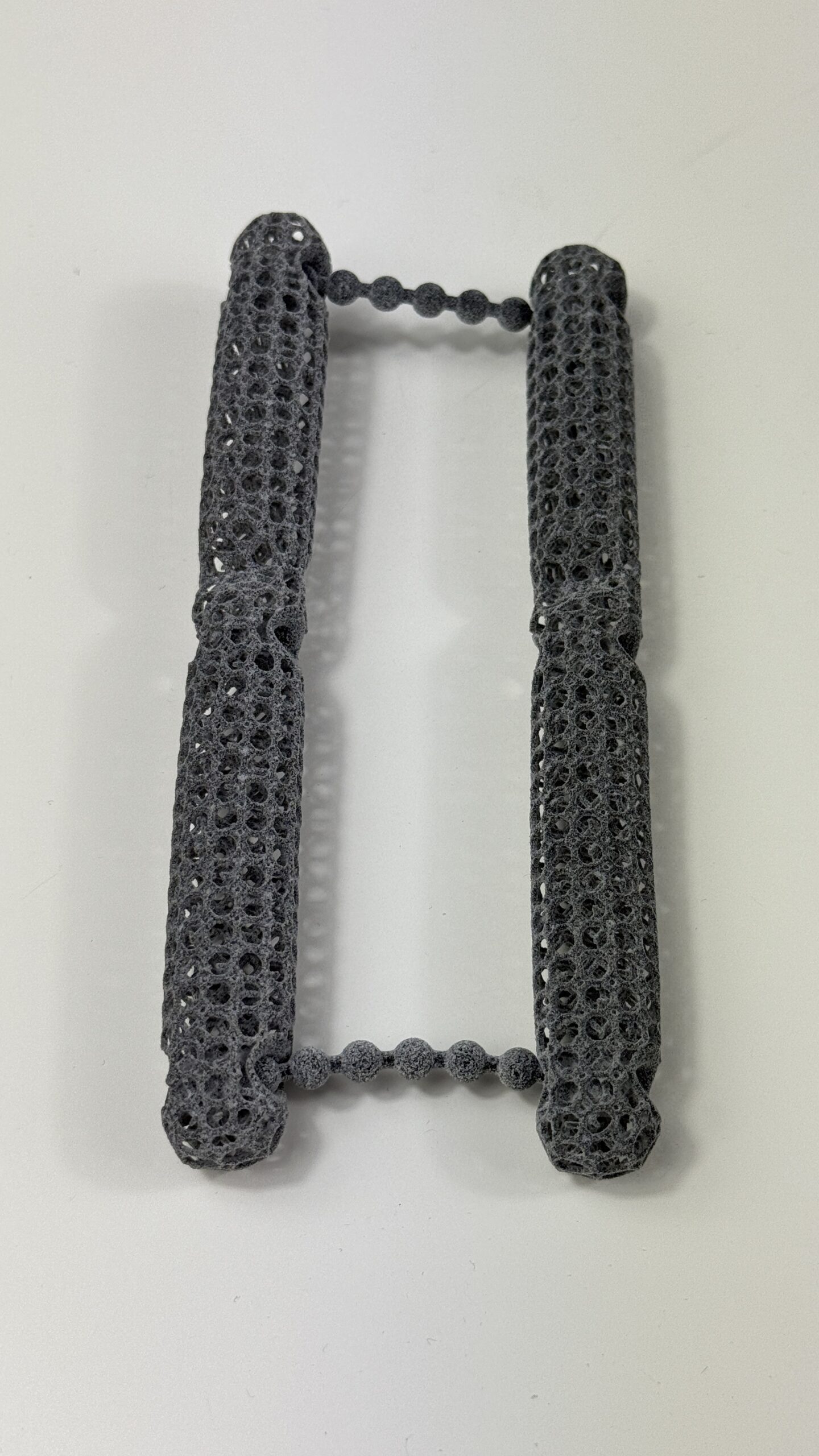
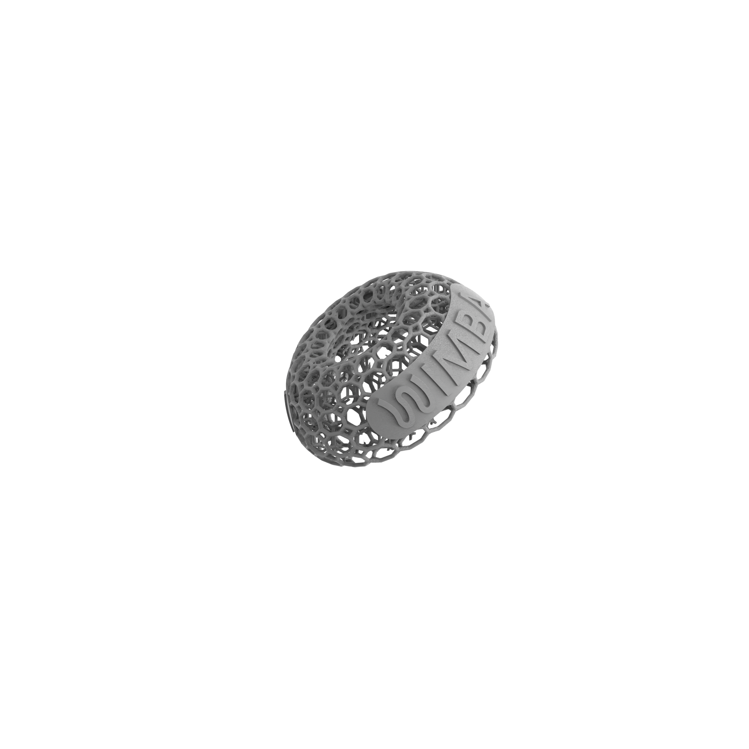
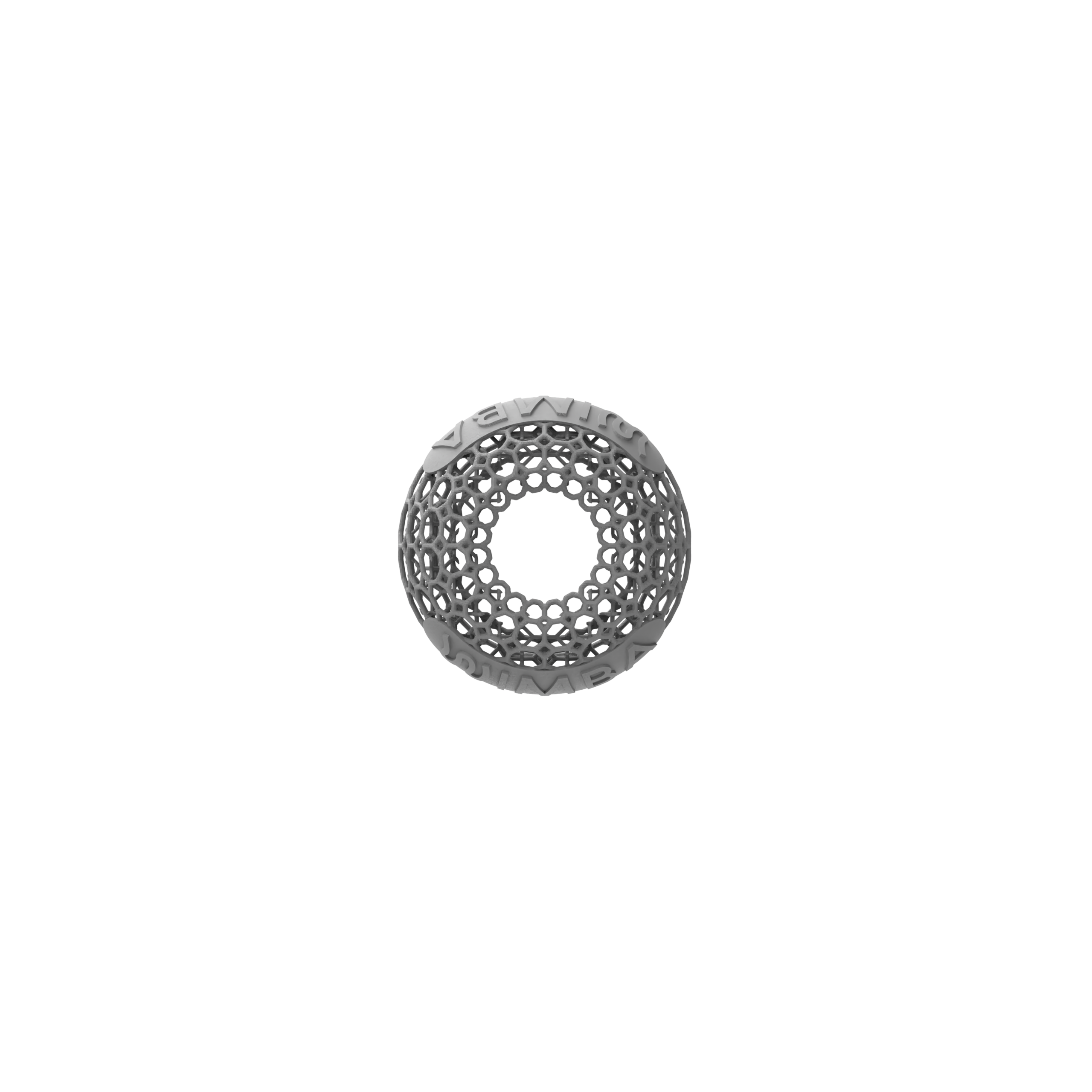
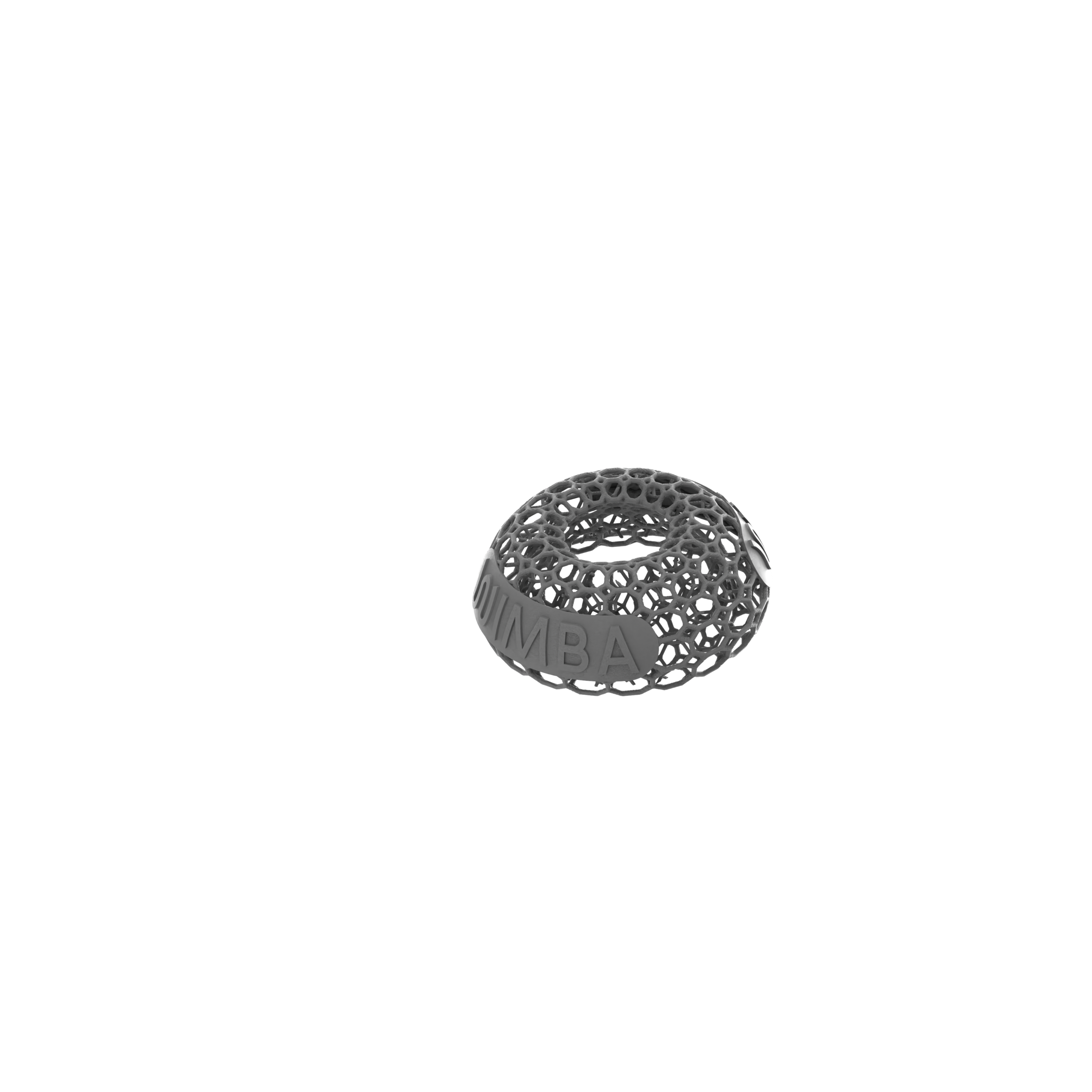
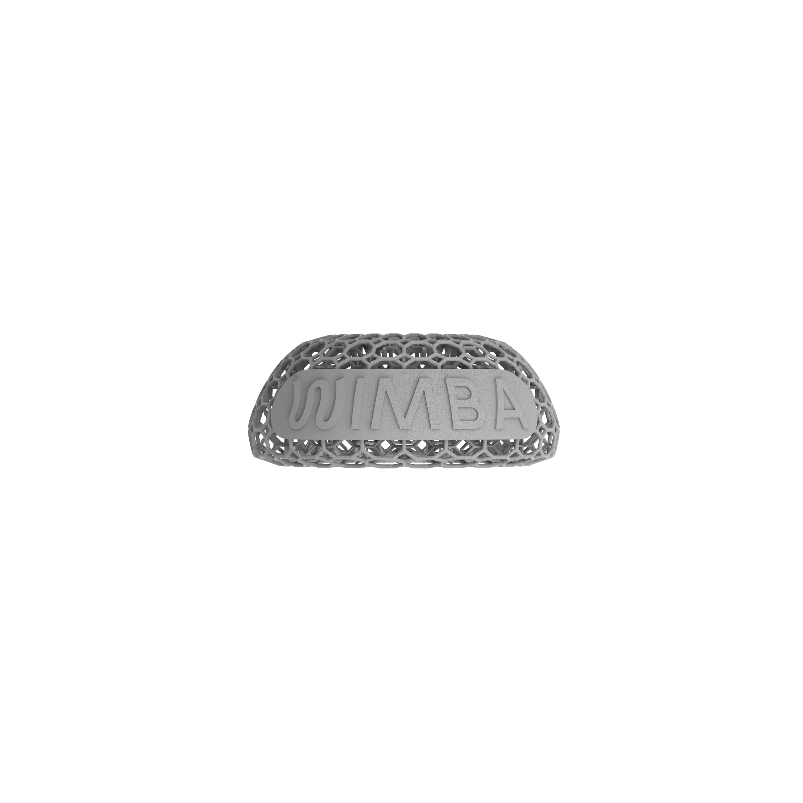
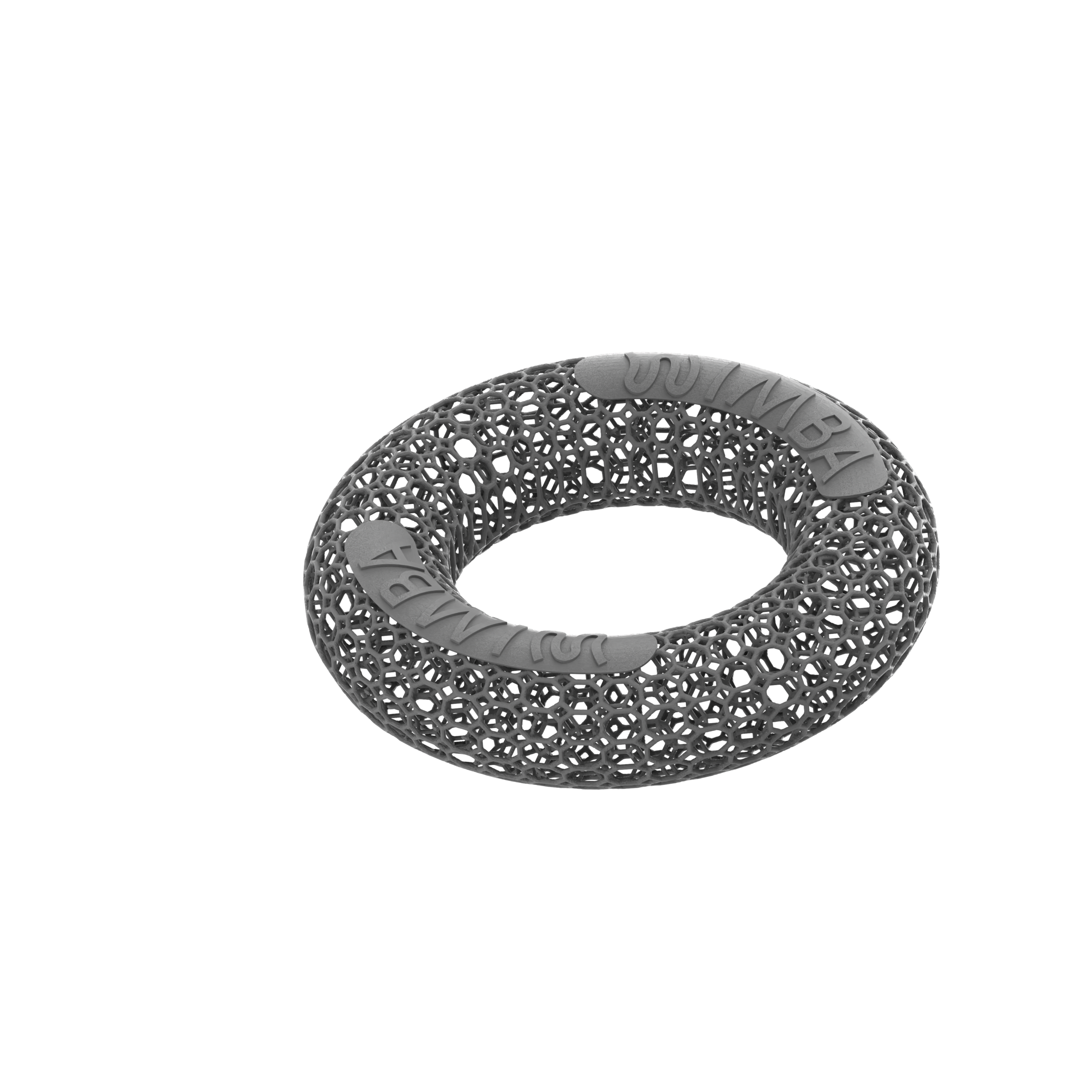
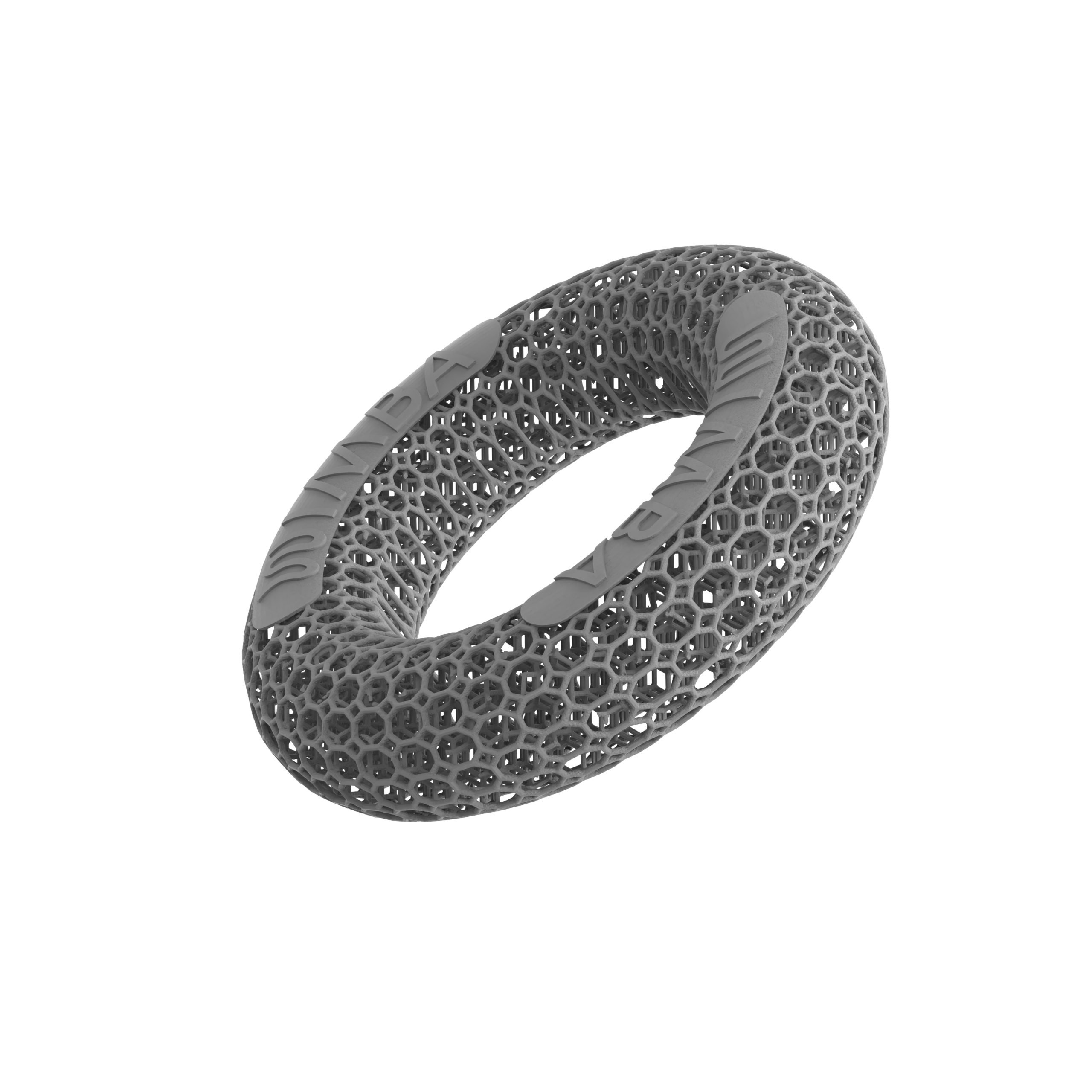
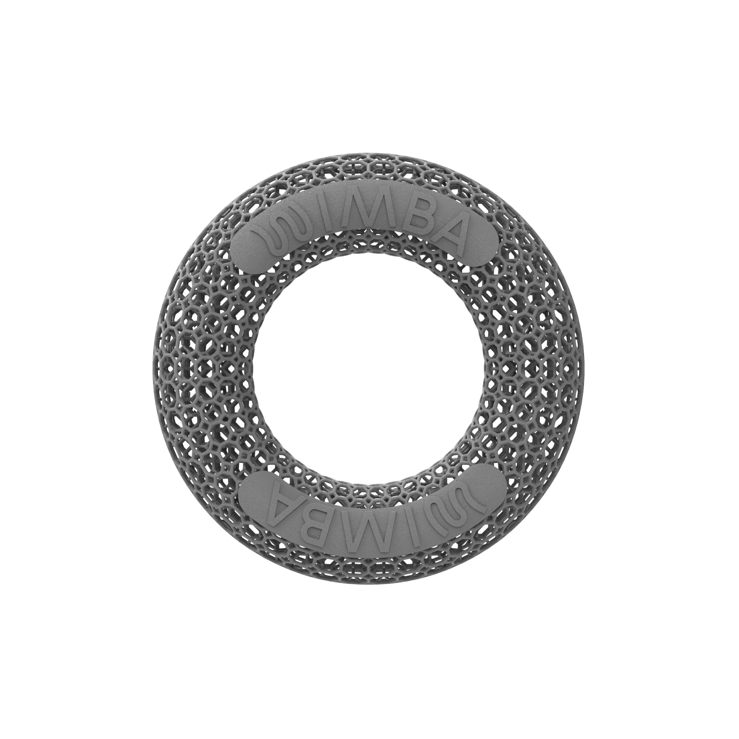
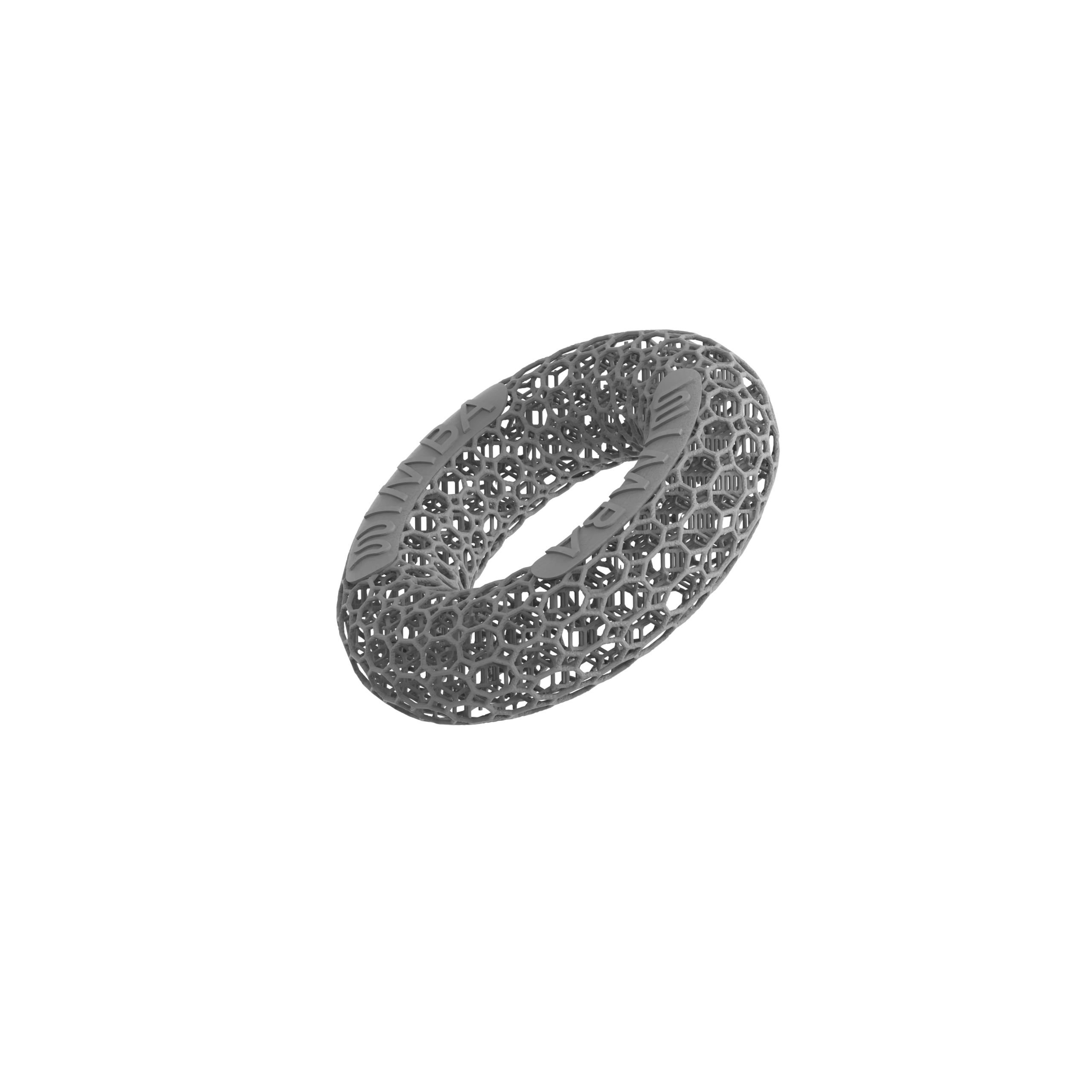
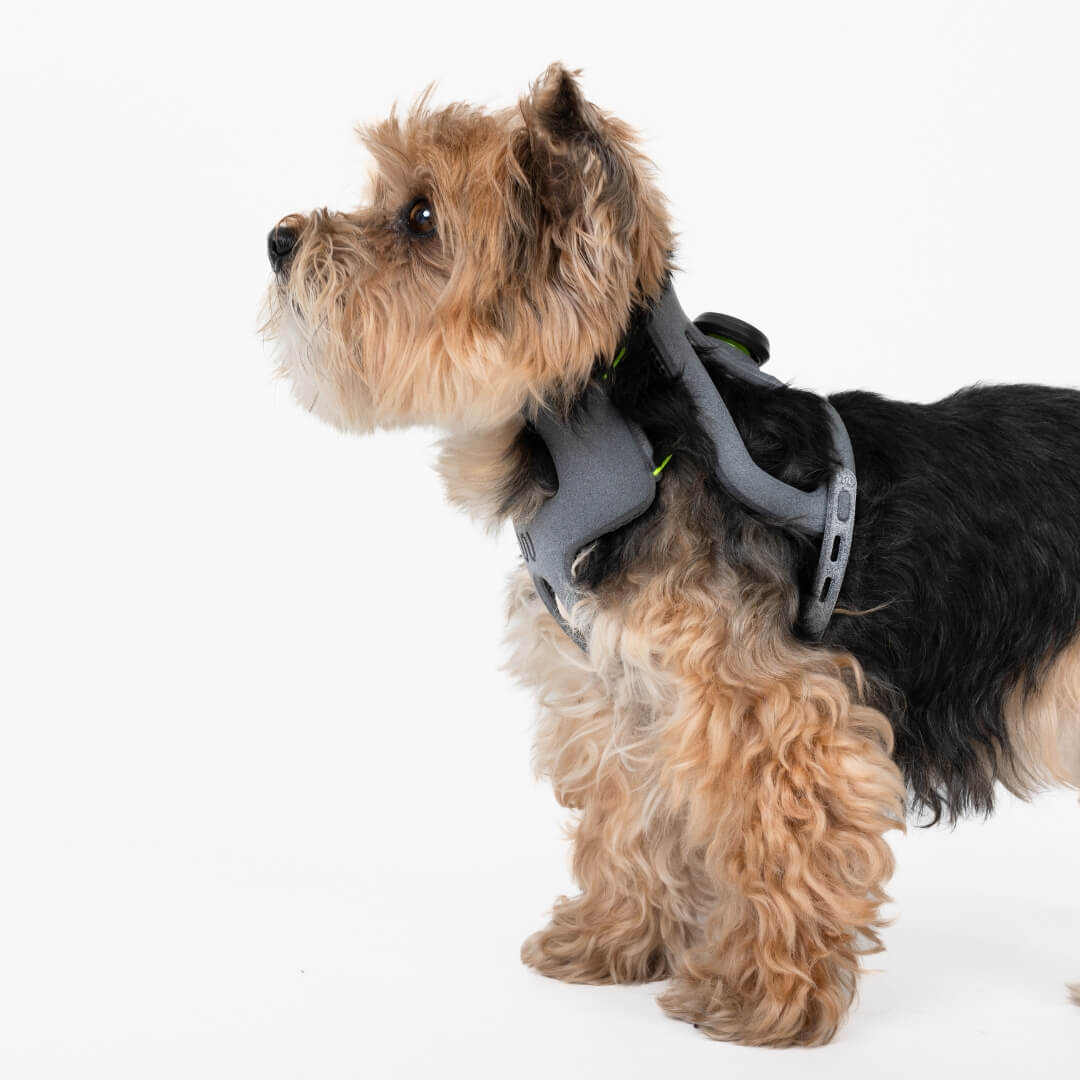
For selected cases of atlantoaxial instability in dogs, WIMBA Cervical Collar GO is an adjustable, 3D-printed external cervical support available for mini and toy-breed C1-C2 patients. It is available in five predefined sizes: Nano, XXS Short, XXS, XS, and S. A veterinary professional takes three measurements, enters them in WimbaAPP, reviews the suggested size, and confirms the final selection. No WimbaSCAN or WimbaBOX is used.
This corrects an important misconception: WIMBA Cervical Collar GO is not individually 3D-scanned or designed from a unique digital model of each dog’s neck. It is a measurement-selected and adjustable device. “3D-printed” describes how the collar is manufactured, not evidence that it will fit every patient or that it is clinically appropriate for every case of atlantoaxial instability in dogs.
For atlantoaxial instability in dogs, the collar may be considered only after neurological assessment and appropriate diagnostic imaging have established the condition and the treating team has defined the goal of external support. It does not replace surgical stabilization when surgery is indicated. Current product information, size limitations, application guidance, and the latest clinical workflow are available on the WIMBA Cervical Collar GO page.
A Real WIMBA Cervical Support Case
WIMBA currently features one cervical patient with a dedicated case-study page: Kacperek. His diagnosis was a severe traumatic cervical spine injury, not atlantoaxial instability in dogs. The case is included as a related example of how external cervical support may be incorporated into a broader veterinary treatment plan. It is an observational patient story, not controlled evidence and not a prediction of outcome for a dog with AAI.
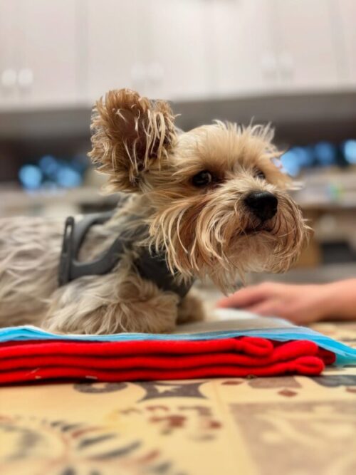
Kacperek: conservative support after severe cervical trauma
Kacperek, a 3.5-year-old Yorkshire Terrier, sustained a crushing cervical injury caused by a larger dog. MRI identified extensive occipital and cervical trauma, spinal cord compression, and secondary myelopathy. Because of the severity of the lesions and the high surgical risk, his team selected conservative stabilization, pain management, strict movement restriction, rehabilitation, and WIMBA Cervical Collar GO. Kacperek progressed from tetraplegia and complete immobility to independent movement. Because several treatments were used together, the improvement cannot be attributed to the collar alone. This was not a case of atlantoaxial instability in dogs.
Home Care After a Diagnosis of Atlantoaxial Instability in Dogs
Home management must follow the treating veterinarian’s written plan. The appropriate restrictions differ between conservative care, the period before surgery, and postoperative recovery. Do not change brace position, wearing time, medication, or allowed activity because the dog appears more comfortable on one day.
- Prevent uncontrolled movement. Block access to stairs, furniture, jumping, running, and rough play according to the prescribed restriction plan.
- Handle the whole body. When lifting is permitted, support the chest and pelvis and avoid pulling or rotating the head and neck.
- Use neck equipment only as directed. The clinical team may recommend a harness rather than attaching a lead to a neck collar, but the exact equipment and walking allowance are patient-specific.
- Monitor neurological function. Report increased pain, changed gait, weakness, knuckling, inability to stand, altered breathing, or loss of bladder or bowel control.
- Monitor external support. Check the areas and schedule specified by the fitting professional. Do not add padding, cut components, or adjust the fitted position without instruction.
- Attend rechecks. Improvement in pain does not prove that C1-C2 stability or spinal cord compression has normalized.
A dog receiving care for atlantoaxial instability may look brighter before the underlying mechanical risk has resolved. Activity should therefore progress according to clinical reassessment and imaging when indicated, not simply because the dog wants to move more.
Prognosis and Follow-Up for Canine Atlantoaxial Instability
Published reports on atlantoaxial instability in dogs document meaningful neurological improvement after both selected nonsurgical management and several surgical techniques, but the evidence is heterogeneous and mostly retrospective.1112 Prognosis cannot be estimated accurately from breed or the presence of a dens abnormality alone.
Important considerations include whether the dog is ambulatory, the duration and progression of signs, the extent of spinal cord injury, mechanical alignment, concurrent craniocervical abnormalities, perioperative risk, complications, and response to initial management. In the 49-dog surgical series, nonambulatory status before surgery was associated with incomplete or absent recovery, while the nonsurgical series found shorter duration of signs associated with a better long-term outcome.1112
Follow-up should therefore assess pain, neurological function, mobility, external device fit when used, and imaging when indicated. A stable-looking dog still requires the planned restrictions and reassessment because atlantoaxial instability in dogs is a structural and neurological condition, not a symptom that can be declared resolved from behavior alone.
Frequently Asked Questions
What is atlantoaxial instability in dogs?
Atlantoaxial instability is inadequate stability between the atlas (C1) and axis (C2), the first two neck vertebrae. Abnormal movement or subluxation can compress or injure the upper cervical spinal cord. Causes include developmental abnormalities of the dens, ligaments, atlas, or wider craniocervical junction, as well as traumatic fracture or ligament disruption.
Which signs of AAI require emergency care?
Inability to stand or walk, rapidly worsening weakness, severe neck pain, collapse, altered consciousness, or difficulty breathing requires emergency veterinary care. Minimize neck movement, contact the receiving clinic for transport instructions, and do not test walking, flex the neck, massage the area, or apply a homemade rigid support.
Can atlantoaxial instability in dogs be treated without surgery?
Some selected dogs can have favorable outcomes with strict activity restriction, prescribed medical care, and professionally managed external cervical support. Evidence is limited to small retrospective series, and recurrence or deterioration remains possible because external support does not create guaranteed permanent fusion. Neurological findings, imaging, progression, anatomy, and patient risk should guide the decision.
How is atlantoaxial instability in dogs diagnosed?
Veterinarians combine the history and neurological examination with carefully planned imaging. Neutral or selected flexed radiographs may assess alignment and bony anatomy, while CT provides detailed bone and joint information and MRI evaluates the spinal cord and soft tissues. No single measurement determines the complete diagnosis or treatment pathway.
Does a dog neck brace for AAI replace surgery?
No. A cervical brace is external support intended to help limit unwanted movement within a veterinarian-directed plan. It does not repair abnormal anatomy, decompress the spinal cord, or guarantee permanent C1-C2 stability. It may be considered in selected conservative, preoperative, or postoperative situations, but surgery remains important when internal stabilization is indicated.
Is WIMBA Cervical Collar GO custom-made for each dog?
No. The collar is not individually 3D-scanned or designed from a unique model of the dog’s neck. It is an adjustable, 3D-printed device available in five predefined sizes. A veterinary professional takes three measurements, reviews the WimbaAPP size suggestion, confirms suitability, fits the collar, and defines the monitoring plan.
AAI Requires a Diagnosis Before a Device
If your dog has neck pain, weakness, ataxia, or a confirmed C1-C2 condition, begin with your veterinarian or veterinary neurologist. When diagnostic imaging and the treatment plan identify a suitable role for external cervical support for dogs, a WIMBA Provider can assess whether Cervical Collar GO fits the patient’s size, anatomy, and clinical objective.
Medical disclaimer: This article is for general educational purposes only and is not a substitute for veterinary examination, neurological assessment, diagnostic imaging, or treatment. Suspected atlantoaxial instability can deteriorate rapidly. Do not flex, rotate, massage, or manipulate a painful neck; test the dog’s ability to walk; give human medication; or apply a homemade brace. Seek emergency care for inability to stand, rapidly worsening weakness, severe pain, collapse, altered consciousness, or breathing difficulty.
Scientific References
- Slanina MC. Atlantoaxial instability. Veterinary Clinics of North America: Small Animal Practice. 2016;46(2):265–275. doi:10.1016/j.cvsm.2015.10.005
- Stalin C, Gutierrez-Quintana R, Faller K, Guevar J, Yeamans C, Penderis J. A review of canine atlantoaxial joint subluxation. Veterinary and Comparative Orthopaedics and Traumatology. 2015;28(1):1–8. doi:10.3415/VCOT-14-05-0064
- Dewey CW, Marino DJ, Loughin CA. Craniocervical junction abnormalities in dogs. New Zealand Veterinary Journal. 2013;61(4):202–211. doi:10.1080/00480169.2013.773851
- Planchamp B, Bluteau J, Stoffel MH, Precht C, Schmidli F, Forterre F. Morphometric and functional study of the canine atlantoaxial joint. Research in Veterinary Science. 2020;128:76–85. doi:10.1016/j.rvsc.2019.11.005
- Takahashi F, Hakozaki T, Kouno S, et al. Epidemiological and morphological characteristics of incomplete ossification of the dorsal neural arch of the atlas in dogs with atlantoaxial instability. American Journal of Veterinary Research. 2018;79(10):1079–1086. doi:10.2460/ajvr.79.10.1079
- Waschk MA, Vidondo B, Carrera I, et al. Craniovertebral junction anomalies in small breed dogs with atlantoaxial instability: a multicentre case-control study. Veterinary and Comparative Orthopaedics and Traumatology. 2019;32(1):33–40. doi:10.1055/s-0038-1675797
- Itoh H, Itamoto T, Tani K, et al. Relationship between clinical parameters and malformations in dogs diagnosed with atlanto-axial instability. PLOS ONE. 2024;19(2):e0293363. doi:10.1371/journal.pone.0293363
- Cummings KR, Vilaplana Grosso F, Moore GE, et al. Radiographic indices for the diagnosis of atlantoaxial instability in toy breed dogs. Veterinary Radiology & Ultrasound. 2018;59(6):667–676. doi:10.1111/vru.12668
- White DA, Renberg WC, Roush JK, Hallman MR, Mauler DA, Milliken GA. Flexed radiographic angles for determination of atlantoaxial instability in dogs. Veterinary Surgery. 2019;48(8):1406–1415. doi:10.1111/vsu.13323
- Planchamp B, Forterre F, Vidondo B, et al. Determination of cutoff values on computed tomography and magnetic resonance images for the diagnosis of atlantoaxial instability in small-breed dogs. Veterinary Surgery. 2022;51(4):620–630. doi:10.1111/vsu.13799
- Havig ME, Cornell KK, Hawthorne JC, McDonnell JJ, Selcer BA. Evaluation of nonsurgical treatment of atlantoaxial subluxation in dogs: 19 cases (1992–2001). Journal of the American Veterinary Medical Association. 2005;227(2):257–262. doi:10.2460/javma.2005.227.257
- Aikawa T, Shibata M, Fujita H. Modified ventral stabilization using positively threaded profile pins and polymethylmethacrylate for atlantoaxial instability in 49 dogs. Veterinary Surgery. 2013;42(6):683–692. doi:10.1111/j.1532-950X.2013.12016.x